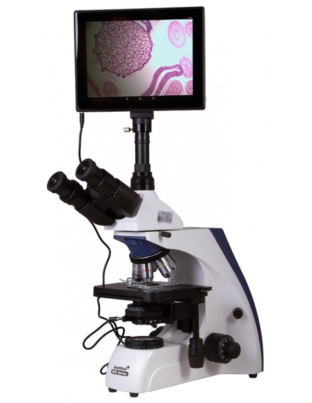
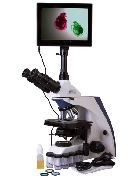
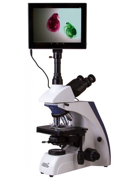
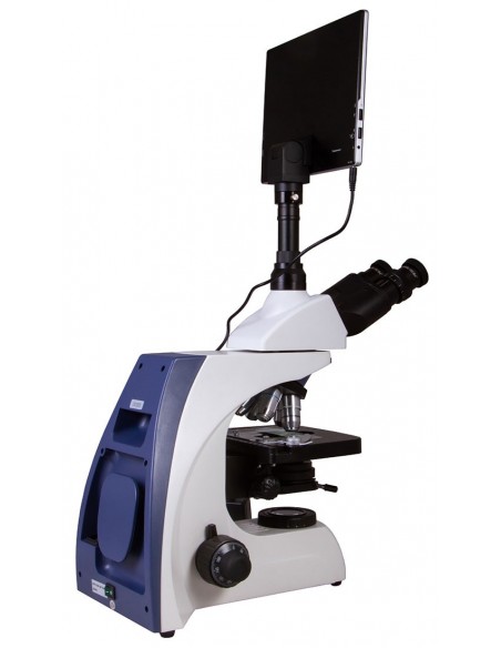
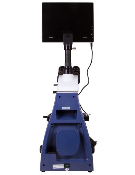
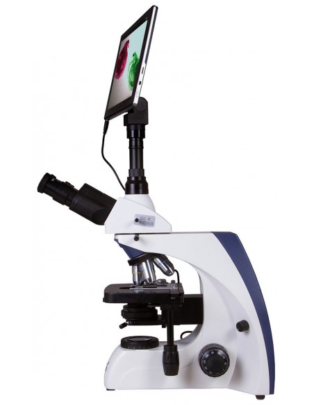
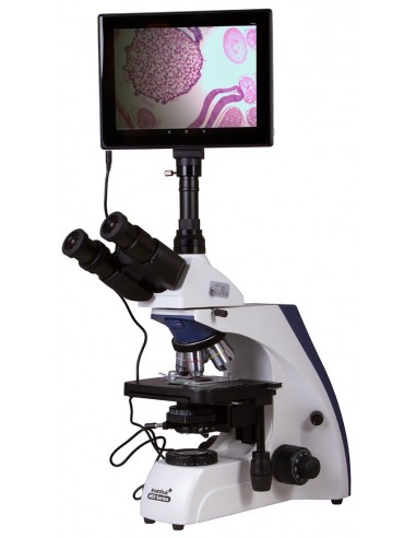
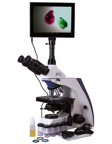
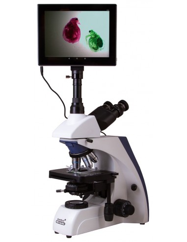
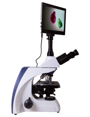
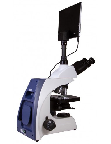
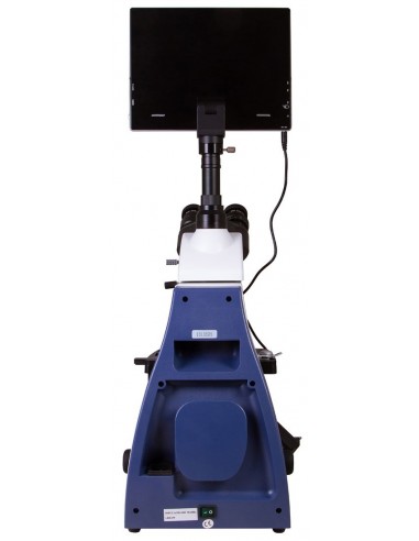
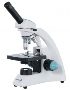
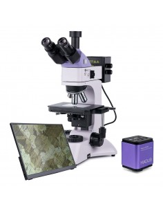
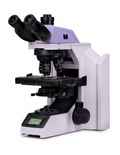
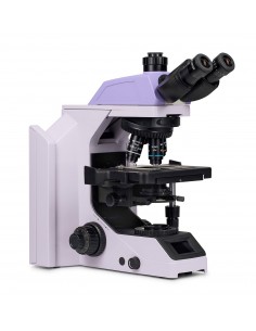
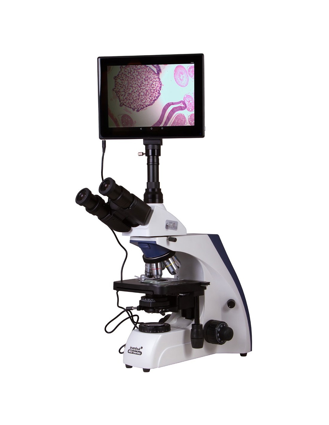
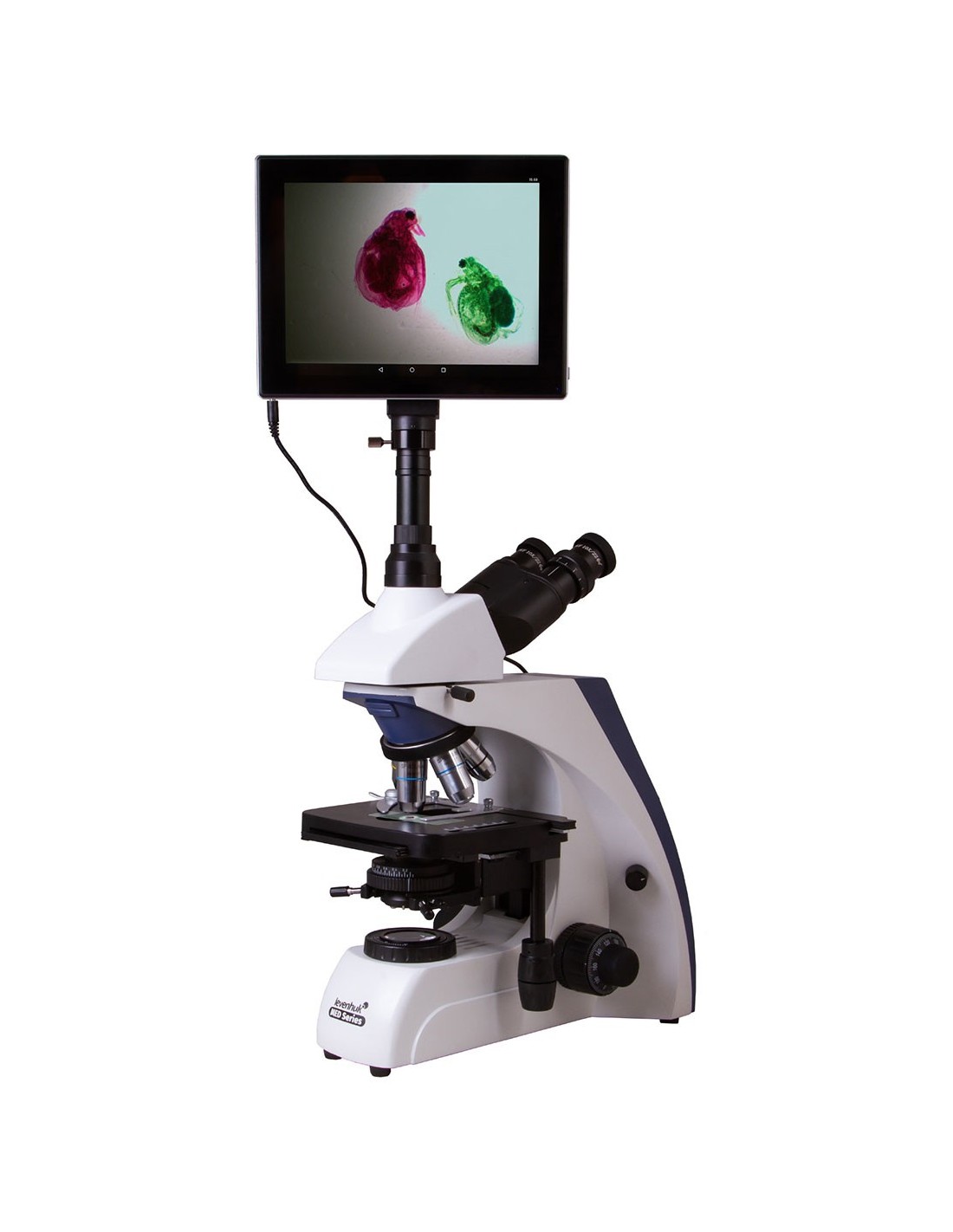
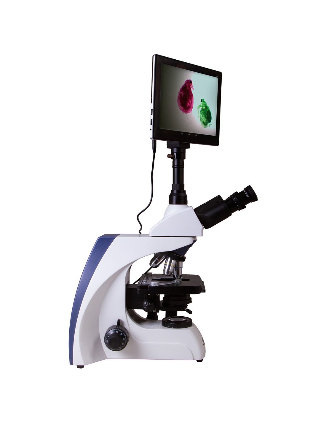
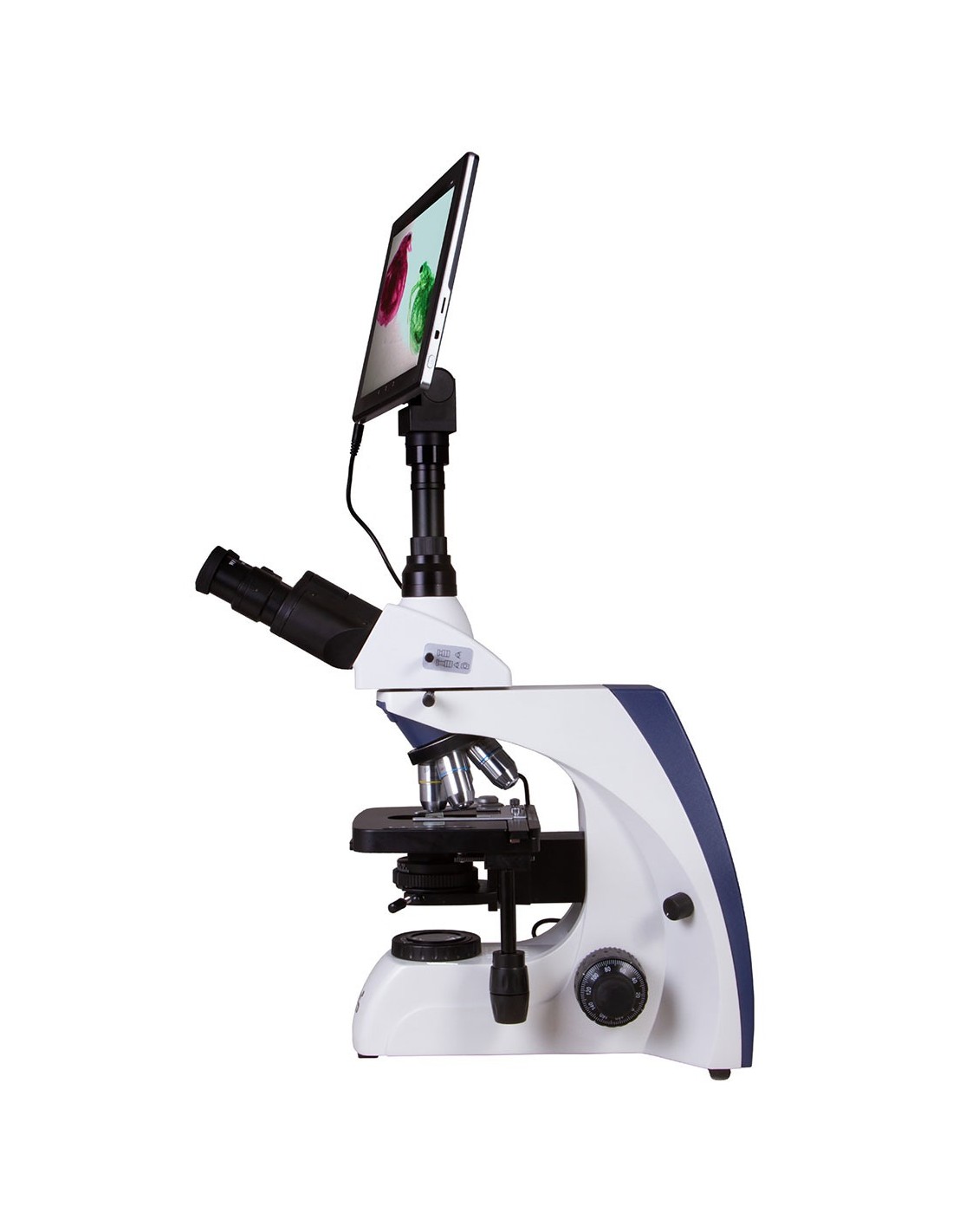
Levenhuk MED D30T LCD is a trinocular digital camera microscope with integrated screen. You don't have to connect the microscope to a computer to set video parameters, transmit sample images to an external device, or process recorded photographs and videos. You can do all this through the camera screen. Furthermore, the microscope has achromatic, semi-planar optical elements with infinite correction and allows you to set the Kohler illumination. The Levenhuk MED D30T LCD is a professional biological microscope suitable for performing various laboratory tasks.
Semi-planachromatic optics with infinity correction
The Levenhuk MED 30 series microscopes are equipped with an infinity corrected optical system, used in professional and high-end microscopes. This system includes semi-planar objectives and allows for clear, high-contrast images with an excellent level of planarity.
The Levenhuk MED 30 are laboratory microscopes (professional, biological) that allow the installation of additional equipment.
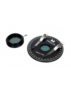
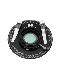
One of the most important features of the infinity corrected optical system is that it allows the installation of additional parts in the optical path between the objective lens and the eyepiece. Additional parts include polarizers (all Levenhuk MED 30 microscopes are equipped with a special slot for their installation), epifluorescence lights and devices. Overall, the modularity and ease of use make the Levenhuk MED 30 microscopes optimal for use in various fields of microscopy, for work in hematology, histology, microbiology and other laboratories.
Rotating head with beam splitter
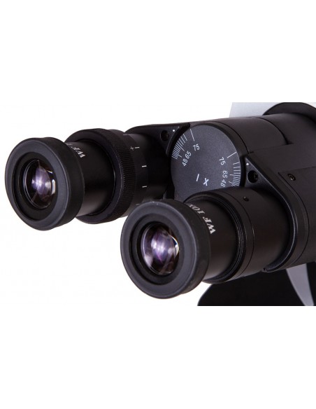
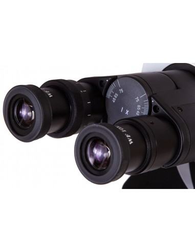
A trinocular head is divided into a viewing part and a tube for installing a digital camera. This allows you to conduct observations through the eyepiece and record at the same time. The head rotates 360º on its axis, which will be appreciated by those who often carry out research in groups. The visual part has an inclination angle of 30º. There is a beam splitter.
Optical glass with anti-mold treatment
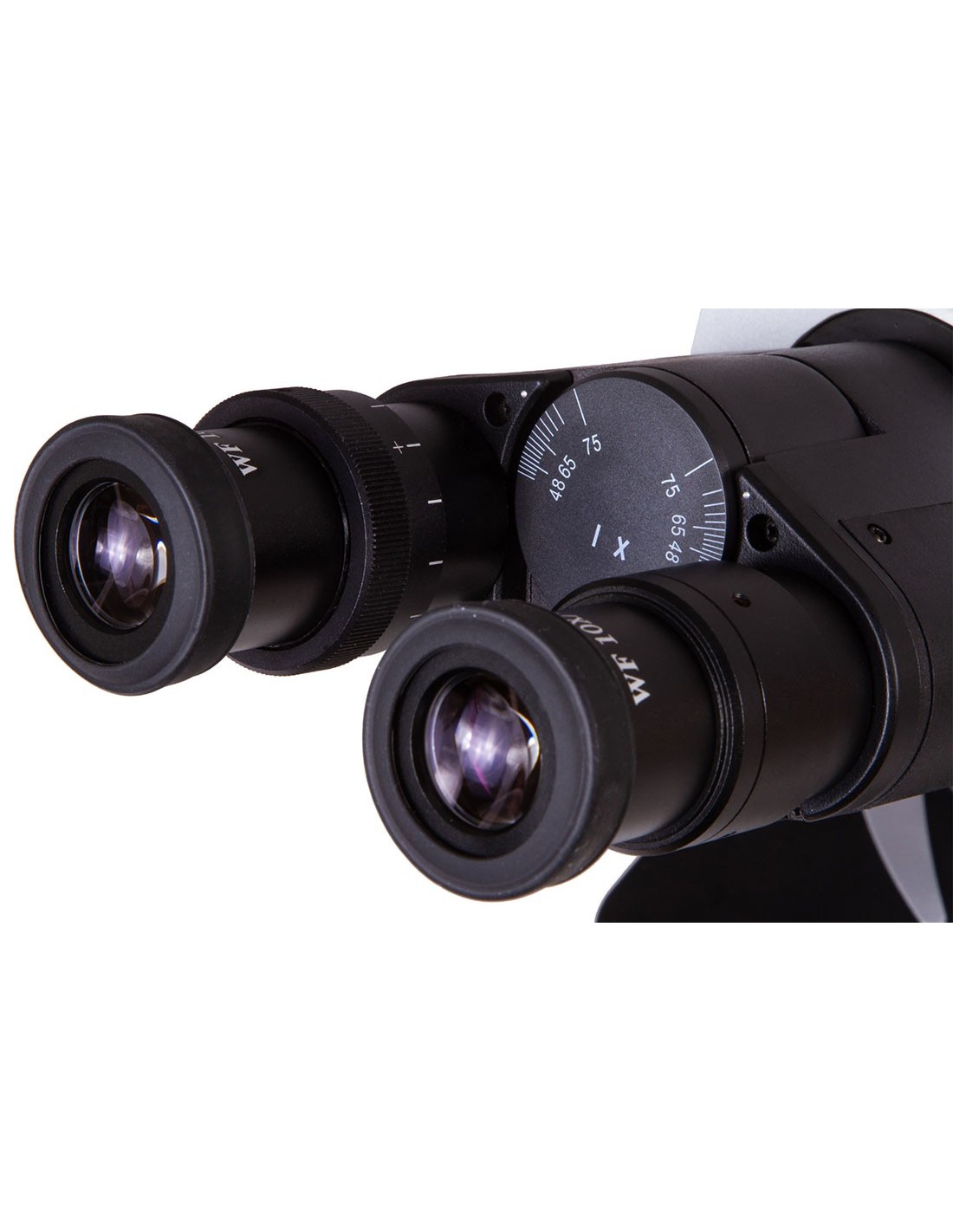
The eyepieces and objective lenses are made of optical glass and protected with an anti-fungal coating. Semi-planar achromatic optical elements flatten the field of view, improve color rendering and reduce chromatic aberrations. The eyepieces offer good vision and 10x magnification. There are five objective lenses. Higher magnification objectives have a protective retractable spring mechanism. A 100x objective lens supports the use of oil immersion. Sharpness adjustment is done by rotating the coarse and fine adjustment knobs.
Digital capabilities
The digital camera does not require the installation of any additional software . You can use any camera option from an LCD screen and see how the micro searches go. You can connect the camera to external displays; It is compatible with several additional accessories (headphones, memory cards and more). A 5MP sensor creates high-quality images and allows you to take high-resolution photographs and record smooth videos at a high frame rate. The software allows you to change image size, brightness and contrast, exposure time and white balance, calibrate the camera and objective lenses, as well as measure specimens and structures (various units of measurement available). A digital camera makes laboratory work easier and more comfortable, as the image is observed on the integrated LCD screen, with touch controls, instead of through the eyepieces, reducing visual stress significantly.
Conveniently work with slides
Under the optical elements, there is a small table with a mechanical scale. Under the coffee table, there is an Abbe condenser with an iris diaphragm. And below that, an LED light with brightness adjustment. You can also set the Kohler lighting. The light is powered by an alternating current source.
The Levenhuk MED 30 are laboratory (biological, professional) microscopes designed for researchers specializing in various fields.
Features :
- Trinocular head with beam splitter, magnification range from 40x to 1000x
- Achromatic, semi-planar optical elements with infinite correction
- The eyepieces and objective lenses have an anti-fungus coating
- Augmented light with 3W LED, with brightness adjustment
- Köhler illumination available
- Powered by an AC source
- The kit includes a 5 MP digital camera and an LCD screen with Android operating system
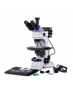
The kit includes:
- Microscope base with support
- 360º rotatable trinocular head
- Achromatic, semi-flat infinitely corrected optical lenses 4x, 10x, 40xs, 60xs, 100xs (oil) with anti-fungal coating
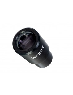
- Wide field eyepieces: CA 10x/22 mm with anti-fungal coating (2 pieces)
- Abbe condenser NA 1.25 with an iris diaphragm
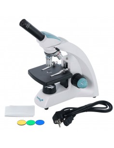
- Filters: blue, green, yellow
- Bottle of immersion oil
- Fuse (2 pcs.)
- Power cable for microscope
- Dust Cover
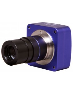
- 5MP Digital Camera with LCD Screen
- Camera Power Cable
- User Manual and Lifetime Warranty
Attention:
Refer to the specification table for the correct mains voltage and never attempt to plug a 110V device into the 220V socket and vice versa, without using a converter. Remember that the mains voltage in the United States and Canada is 110 V and 220–240 V in most European countries.
Some things you can observe under a microscope:
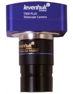
The Levenhuk MED D30T LCD digital trinocular microscope is compatible with Levenhuk digital cameras (purchased separately). Levenhuk cameras are installed in the eyepiece tube, in place of an eyepiece.
| Brand | Levenhuk Inc., USA |
| Lifetime | guarantee |
| EAN | 5905555005010 |
| Package size (LxWxH) | 62x46x28 cm |
| Shipping weight | 11.54 kg |
| Type | digital, light/optical, biological |
| Trinocular | head | td> |
| Optics material | optical glass with anti-mold coating |
| Vertical tube | 360° rotatable, with light beam switch (splitter) |
| Head inclination angle | 30° |
| Magnification, x | 40 — 1000 |
| Magnification range | 800x to 1280x |
| Ocular tube diameter, mm | 30 mm (binocular head), 23.2 mm (third vertical tube) |
| WF10x/22 mm, wide field with diopter adjustment (2 pcs.) |
| Objectives | aromatic optical lenses , semi-flat infinite correction 4x, 10x, 40xs, 60xs, 100xs (oil) |
| Nosepiece nosepiece | for 5 objectives |
| Interpupillary distance, mm | 48 — 75 |
| Stage, mm | 180x160 |
| Range of movement of the stage, mm | 80/50 |
| Stage characteristics | mechanical double layer |
| Eyepiece diopter adjustment, diopters | ±5 |
| Condenser< /td> | Abbe condenser with NA 1.25 and iris diaphragm |
| Diaphragm | iris |
| Fire | coaxial, coarse (0.5 mm) and fine (0.002 mm), with rack and pinion |
| Body | metal |
| Lighting | LED |
| Brightness adjustment | yes |
| Power supply | 100–240V |
| Type of light source | 3 W, with an extra lens |
| Optical filters | blue, green, yellow |
| Extra elements | manifold, Köhler lighting |
| Special features | 9.4-inch color LCD screen, sensor;
integrated memory: 4 GB |
| Possibility to connect additional equipment | support of microSD cards with capacities up to 32 GB;
monitor/TV (with HDMI port);
USB flash drive, computer mouse, keyboard (with a USB connector); headphones (3.5 mm) |
| User level | power users, professionals |
| Professional level difficult assembly and installation | complicated |
| Application | laboratory/medical |
| Lighting position | lower |
| Search method | bright field |
| Digital camera included | yes |
| Set with case/case/bag | dust cover |
| Maximum resolution | 2048x1536 |
| Megapixel | 5 |
| Sensor element | 1/2.5" |
| Pixel size, µm | 2.2x2.2 |
| Video recording | yes |
| Image format | *.jpg |
| Video format | 1080p, *.3gp |
| White balance | manual, automatic |
| Exposure control | manual, automatic |
| Sensitivity, V/lux-sec @550 nm | 0.53 |
| Frame rate | 15 frames per second |
| the third eyepiece tube of the microscope 23.2 mm |
| Software, drivers | Android 5.1 ( multilingual) |
| Programmable options | measurement, brightness, grain size analysis, etc. |
| Output< /td> | USB 2.0 (2 pcs.), mini HDMI, Wi-Fi, TF memory card slot |
| Camera power | DC 12V/2A; via AC adapter |