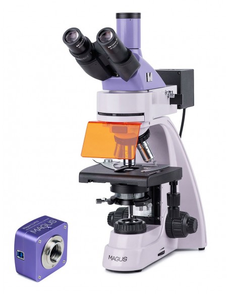
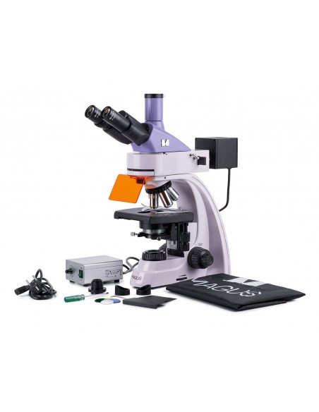
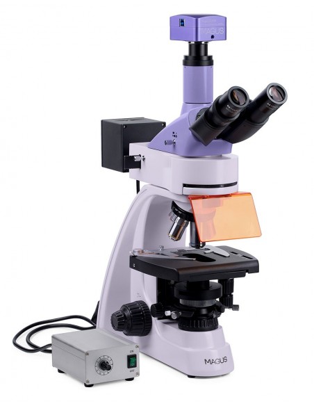
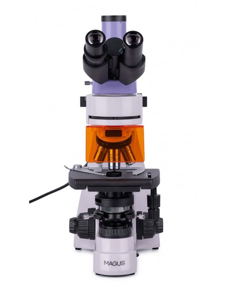
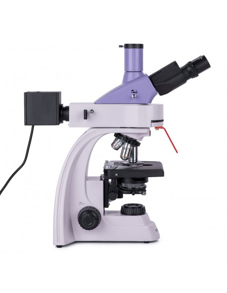
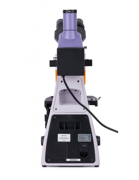
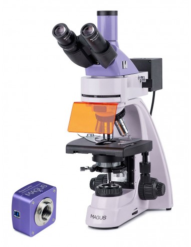
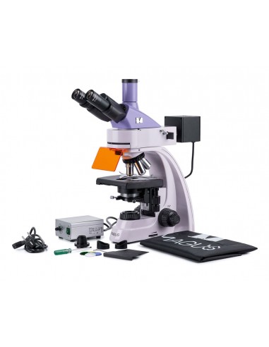
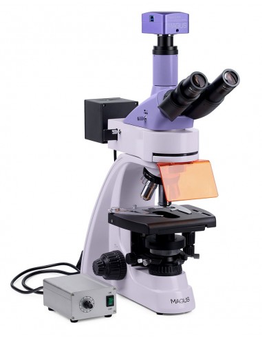
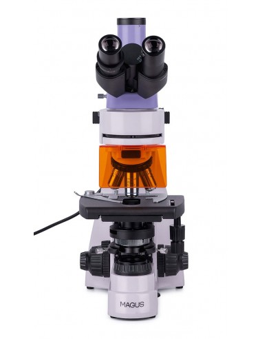
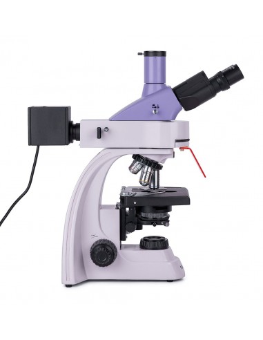
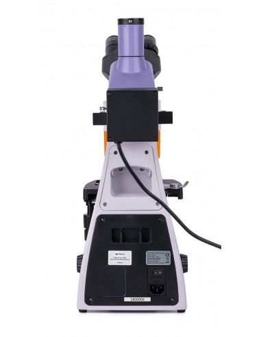
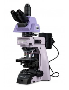
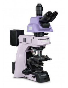
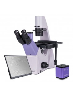
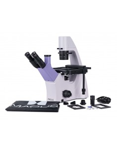
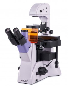
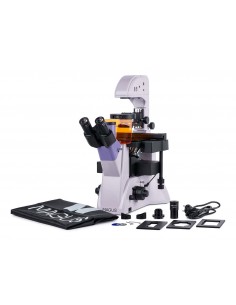
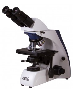
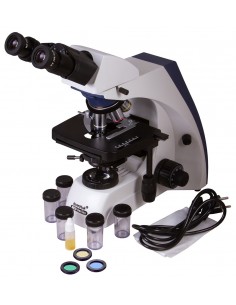
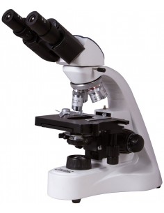
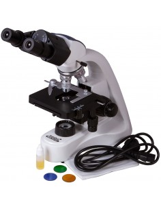
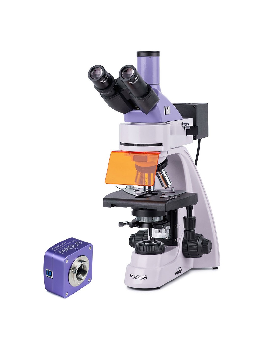
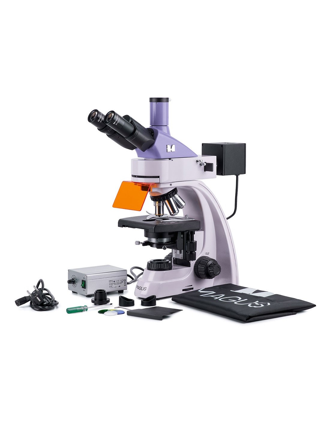
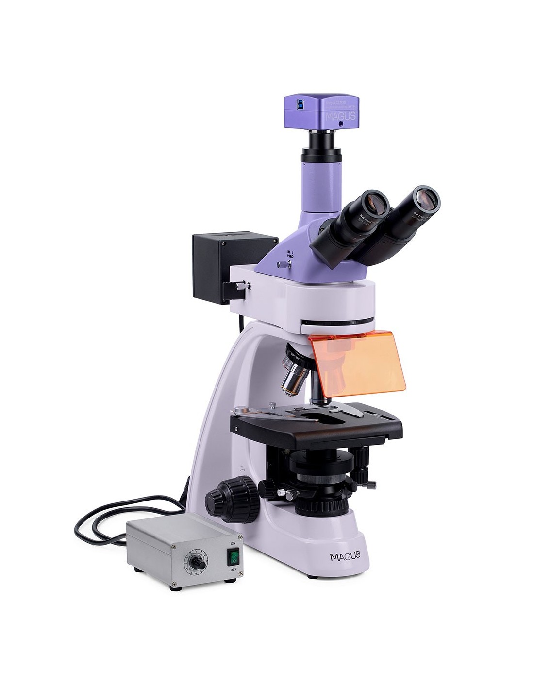
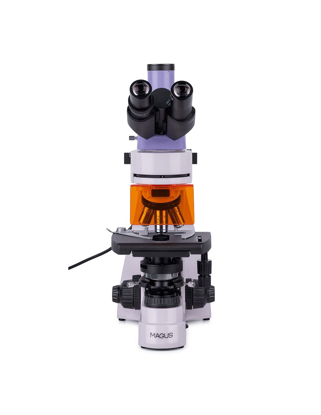
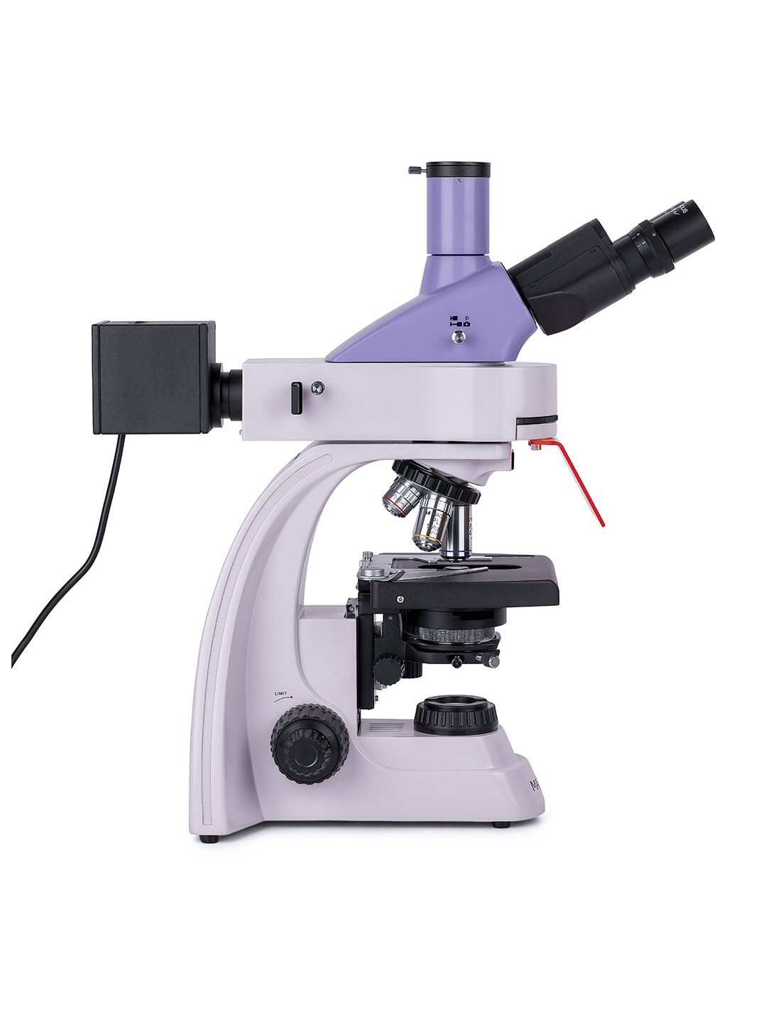
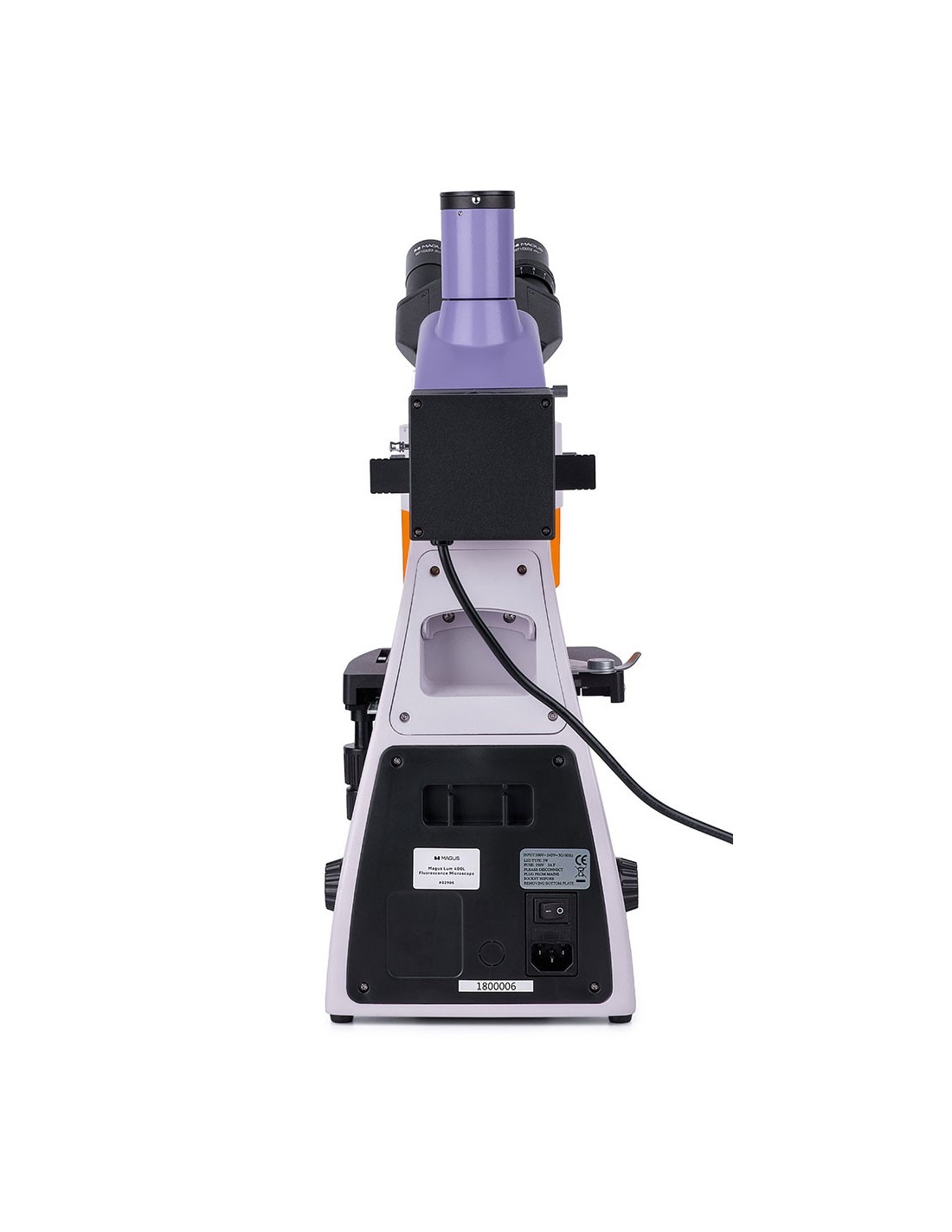
The MAGUS Lum 400L fluorescence microscope can be used for two microscopy techniques: fluorescence in reflected light and bright field in transmitted light. When equipped with additional accessories, the microscope can be used to conduct research in polarized light and for phase contrast and dark field techniques. You can use the microscope for laboratory research and diagnostics in the fields of medicine, pharmacology, forensic science, biotechnology, veterinary medicine, etc.
Digital camera
The MAGUS CLM10 digital camera is designed for dark-field and fluorescence microscopy techniques. The camera is equipped with a 2.3 MP sensor and produces realistic images at a resolution of 1920х1200 px. It is recommended for 40x, 60x and 100x objectives. When making observations with medium magnification lenses, the camera will produce even sharper details.
Videos are recorded at 120 fps at full resolution. Videos flow smoothly, with smooth, subtle transitions between frames. The movement of the object is shown in real time, without delay. The camera facilitates the observation of moving objects and is suitable for academic demonstrations.
The camera is equipped with a USB 3.0 interface. The data transfer speed is 10 times faster than USB 2.0 cameras. The high-speed camera is recommended for professional laboratories, research or university education.
Optics
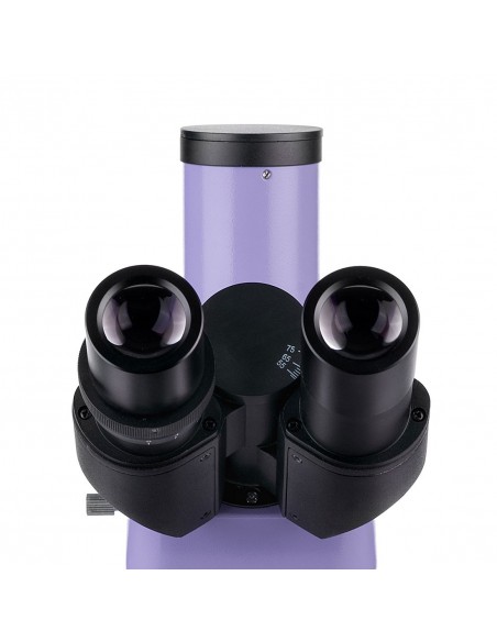
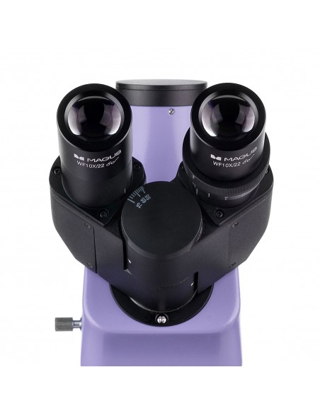
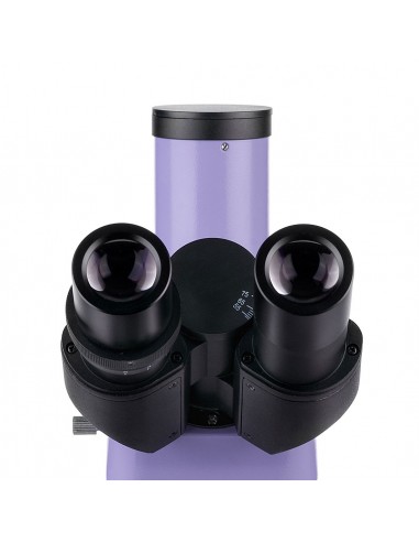
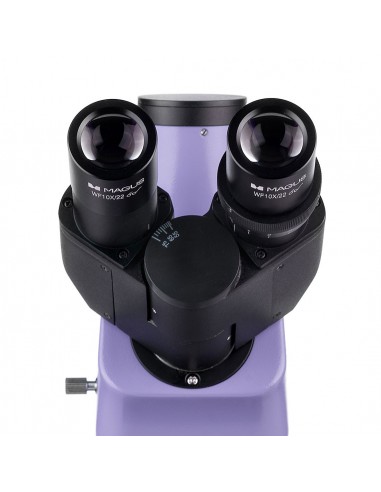
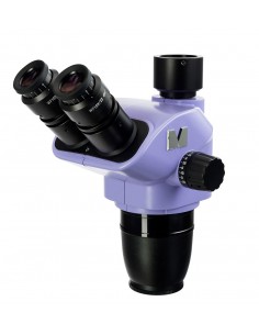
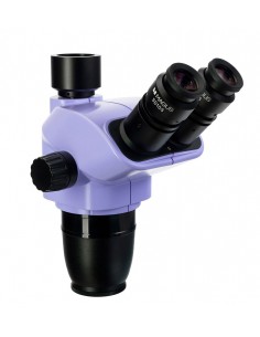
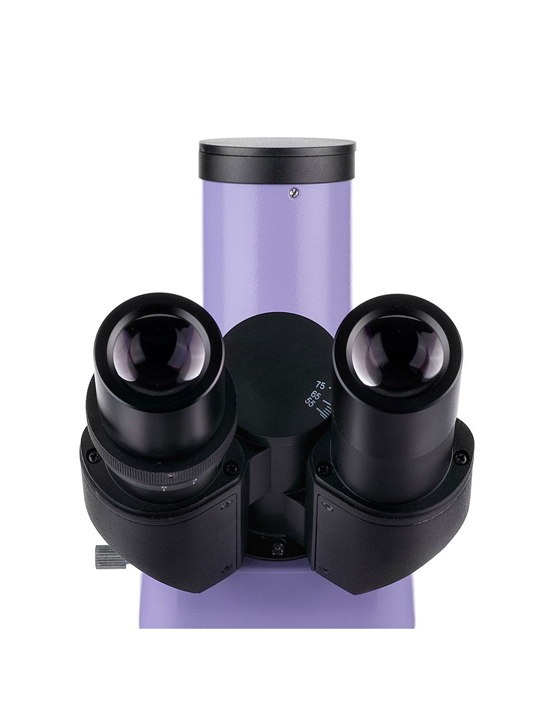
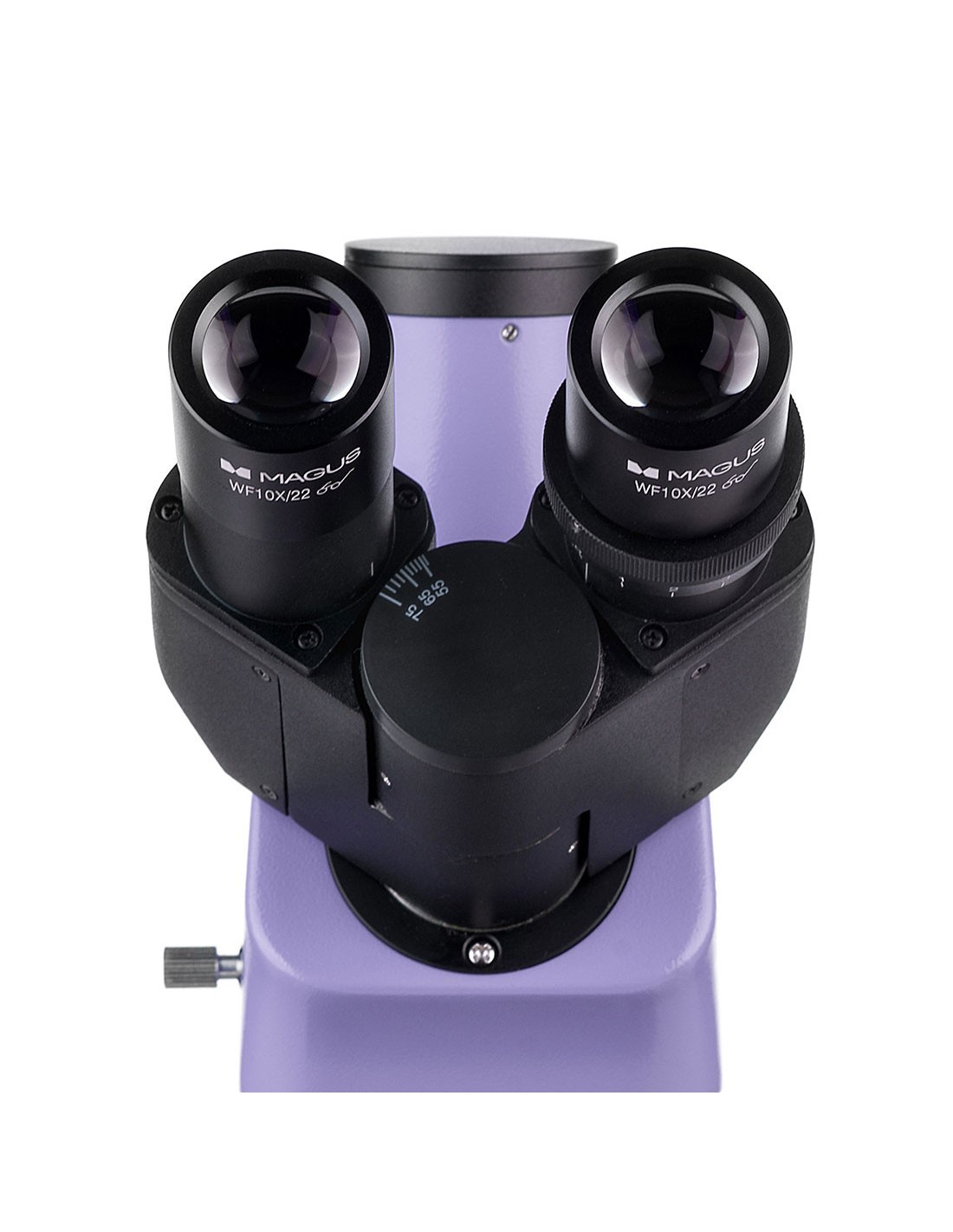
The trinocular head consists of two optical tubes for conducting observations and a trinocular tube for housing a digital camera (not included). To face long periods of observation in maximum comfort, it is possible to adjust the eyepiece tubes by rotating them 360° to adapt to the height of the observer. They are inclined by 30°.
The nosepiece with five objectives is oriented inwards, so that the user can see which objective is inserted in the optical path. It is equipped with infinity planachromatic objectives: three for the bright field technique and one for the fluorescence (fluorescent) technique. Once all objectives are installed on the nosepiece, one slot remains available. It is used to adjust the position of the illuminator for reflected light observations. You can also install an optional lens to achieve higher magnification.
Illumination
Reflected light: five 5W LEDs. Each LED falls into four bands specifications: blue (B), green (G), violet (V) and ultraviolet (UV). To choose the LED you need to rotate the ring. The advantage of LED lighting is that it quickly reaches maximum brightness and turns off quickly, making it easy to switch from brightfield to fluorescence and vice versa. The LEDs do not overheat, do not require a heat sink and have a lifespan of 50,000 hours before needing replacement. LED fluorescence is a safe and easy to maintain microscopy technique, therefore it is recommended for use in schools and universities.
The transmitted light is produced by a 3 W LED. The life cycle of LEDs is 50,000 hours. The lighting system also includes a capacitor. The condenser has an available slot for a dark field slider or a phase contrast slider. The slider makes it quick and easy to switch from one microscopy technique to another without having to change the condenser.
In transmitted and reflected light, Köhler illumination can be set. The study object is sharp and has no image artifacts. The edges do not darken and each objective reaches maximum resolution.
Stage and focusing mechanism
The microscope stage is equipped with a holder removable sample in order to perform manual scans of the same. The table does not have a positioning rack, therefore it requires less working space. You can install a black plate on the stage to eliminate any unwanted light from the transmitted light light source when observing in reflected light.
The coaxial fine (micrometer screw) and coarse (micrometer screw) focus knobs macrometric) are positioned on both sides at the base of the microscope. The coarse focus is equipped with a fixing knob and tension adjustment.
Accessories
The MAGUS Lum 400L microscope features a wide range of compatible optional accessories that can be used to expand its potential. They include eyepieces, objectives, dark field, phase contrast and polarized light devices, digital cameras and calibration slides.
Key Microscope Features:
- Study of reflected light samples using the fluorescence microscopy technique and transmitted light using the bright field microscopy technique.
- Planachromatic objectives at infinity: three bright field objectives and one fluorescent objective
- Trinocular head with 360° rotatable body, trinocular tube for mounting a digital camera
- Four reflected light LEDs with four different wavelengths: blue (B), green (G), violet (V) and ultraviolet (UV
- 3 W LED with transmitted light, Köhler illumination with reflected and transmitted light
- Stage without positioning rack, sample holder removable
- Wide range of accessories, including devices for phase contrast, polarized light and dark field
Key camera features:
- For darkfield and fluorescence microscopy with 40x, 60x and 100x objectives
- 2.3 MP resolution for detailed images at high magnification; low noise level, low power consumption
- 120 fps for observing moving objects, video recording, and moving the sample without delays or stutters
- Global shutter for fast reading of the signal from the sensor, increased image brightness without optical effects during observations of moving objects
- The monochrome back-illuminated SONY Exmor CMOS sensor offers images with low noise and high sensitivity to light even in of low light. You can get sharper, brighter and more saturated color images.
- USB 3.0 interface for fast and smooth data transfer
- Software for recording and editing photos and videos, recording functions external display, linear and angular measurements
The package contains:
- MAGUS CLM10 digital camera (digital camera, USB cable, Installation CD with drivers and software, user manual and warranty certificate)
- Support with integrated power supply, transmitted light source, focusing mechanism, stage, condenser mount and nosepiece
- Abbe condenser
- Illuminator for reflected light observations
- Housing of the LED lamp
- Trinocular head
- Planachromatic objective infinity: PL 4x/0.10 WD 19.8 mm
- Infinity plan achromatic objective: PL 10x/0.25 WD 5.0 mm
- Infinity plan achromatic objective, fluorescent: PL FL 40x/0.85 (with spring mechanism) WD 0.42 mm
- Infinity planachromatic objective: PL 100x/1.25 (with spring mechanism, oil) WD 0 .36 mm
- 10x/22 mm eyepiece with long eye relief (2 pcs.)
- Eyecup (2 pcs.)
- UV screen
- Filter slider
- Black protective plate for installation under the table
- C-mount adapter
- Color filter (4 pcs.)
- Bottle of immersion oil
- Reflected light illuminator power supply
- Power cord
- Reflected light illuminator power cord
- Reflected light illuminator power cord
- Dust cover
- User manual and warranty card
Available upon request:
- 10x/22 mm eyepiece with graduated scale
- 12.5x/14 mm eyepiece (2 pcs.)
- 15x eyepiece /15 mm (2 pcs.)
- Eyepiece 20x/12 mm (2 pcs.)
- Eyepiece 25x/9 mm (2 pcs.)
- Objective infinity planachromatic, fluorescent: PL FL 10x/0.35 WD 2.37 mm
- Infinity planachromatic objective: PL 20х/0.40 WD 8.8 mm
- Infinity planachromatic objective: PL 60x/0.80 ∞/0.17 WD 0.46 mm
- Phase contrast device
- Dark field condenser with NA 0.9
- Oil-filled dark field condenser with NA 1.36–1.25
- Dark field slider
- Polarizer tool
- Calibration glass
- LCD monitor
| Brand | MAGUS |
| EAN | 5905555018188 |
| 45x30x96 cm |
| Shipping weight | 15.1 kg |
| === Delimiter === | Microscope specifications |
| Type | biological, light/optical, digital |
| Tested | trinocular |
| Vertical tube | Gemel head (Siedentopf type, with 360° rotation) |
| Head inclination angle | 30° |
| Eyepiece tube diameter, mm | 30 |
| Eyepieces | 10х/22 mm, eye relief: 10 mm (*optional: 10x/22 mm with graduated scale, 12.5x/14; 15x/15; 20x/12; 25x/9) |
| Objectives | infinity planachromatic and fluorescent objectives: PL 4x/0.10, PL 10x/0.25, PL FL 40x/0.85, PL 100x/1.25 (oil); parfocal distance 45 mm (*optional: PL FL 10x/0.35, PL 20х/0.40, PL 60x/0.80 ∞/0.17) |
| Revolver nosepiece | for 5 objectives |
| Working distance, mm | 19.8 (4x); 5.0 (10x); 0.42 (FL 40x); 0.36 (100x); 2.37 (FL 10х); ); 8.8 (20х); 0.46 (60х) |
| Interpupillary distance, mm | 48 — 75 |
| Stage, mm | 180x150 |
| Range of movement of the stage, mm | 75/50 |
| Stage characteristics | two-axis mechanical translator, without positioning rack |
| Condenser | condenser from Abbe, NA 1.25, adjustable centering, adjustable height, adjustable aperture diaphragm, with slot for dark field slider and phase contrast slider, dovetail mount |
| Diaphragm< /td> | adjustable aperture diaphragm, field diaphragm with adjustable iris |
| Focus | coaxial, coarse focus (21 mm, 39 .8 mm/turn, with fixing knob and micrometer screw tension adjustment knob) and fine focus (0.002 mm micrometer screw) |
| Illumination | LED, fluorescent |
| Brightness adjustment | yes |
| Power supply | 85–265 V, 50/60 Hz |
| Type of light source | reflected light: 4 LEDs of different wavelengths , 5W; transmitted light: 3 W LED |
| Optical filters | yes |
| Possibility to connect equipment additional | phase contrast devices (condenser and objectives), dark field condenser (dry or oil immersion), polarization instruments (polarizer and analyzer), dark field slider |
| User level | expert users, professionals |
| Assembly and installation level of difficulty | complicated |
| Fluorescent module | filters: ultraviolet (UV), violet (V), blue (B), green (G) |
| Fluorescence filter: filter type/excitation wavelength/dichroic mirror/emission wavelength | ultraviolet (UV), 320–380 nm/ 420nm/435nm; violet (V), 380–415 nm/460 nm/475 nm; blue (B) 410–490 nm/505 nm/515 nm; green (G) 475–550 nm/580 nm/595 nm |
| Application | laboratory/medical |
| Illumination position | double |
| Research method | bright field, fluorescence |
| === Delimiter === | Digital camera |
| Sensor | SONY Exmor CMOS |
| Color/monochrome |
| Megapixel | 2.3 |
| Maximum resolution, px | 1920х1200 |
| Sensor size | 1/1.2" (11.25x7.03 mm) |
| Pixel size, µm | 5.86x5.86 |
| Light sensitivity | 1016 mV with 1/30 s< /td> |
| Signal-to-noise ratio | 0.15 mV at 1/30 s |
| Exposure time | 0.244 ms – 2 s |
| Video recording | yes |
| Video recording | yes |
| Frame rate, fps@resolution | 120@1920x1200 |
| Installation location | trinocular tube , optical tube instead of eyepiece |
| Image format | *.jpg, *.bmp, *.png, *.tif |
| Video format | recording: *.wmv, *.avi, *.h264 (Windows 8 and later), *h265 (Windows 10 and later) |
| Spectral range, nm | 380–650 (with IR filter and anti-reflection filter) |
| Shutter type | Global shutter |
| White balance | manual, automatic |
| Exposure control< /td> | manual, automatic |
| Software features | image size, brightness, exposure control |
| Output | USB 3.0, 5 Gb/s |
| System Requirements | Windows 8/10/11 (32-bit and 64-bit), Mac OS minimum 2 GB RAM, USB 3.0 port, CD-ROM, 17" or larger |
| Mount type | C-mount |
| Body | CNC aluminum alloy |
| Camera power supply | DC, 5V, from the computer's USB port; 12 V, 3 A adapter for the Peltier cell |
| Camera operating temperature range, °C | -10 — 50 |
| Humidity operating range, % | 30 — 80 |