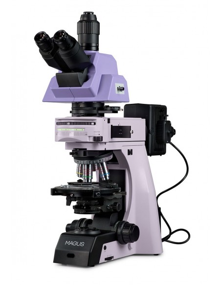
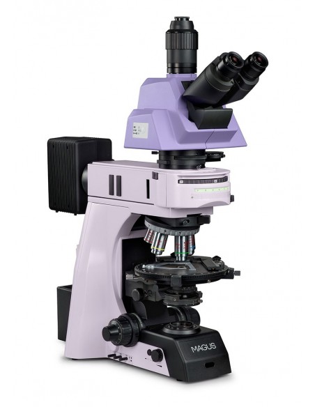
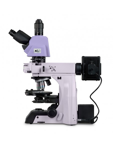
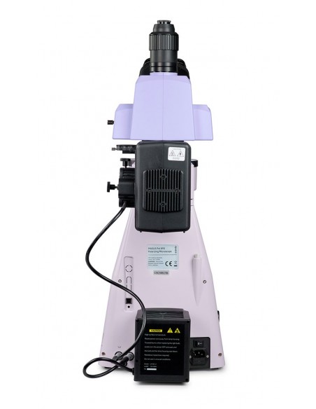
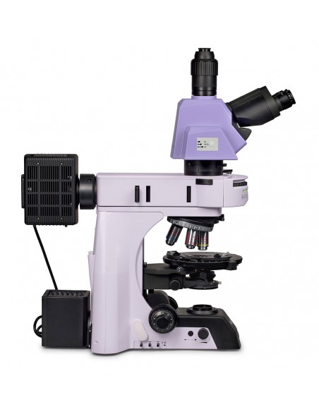
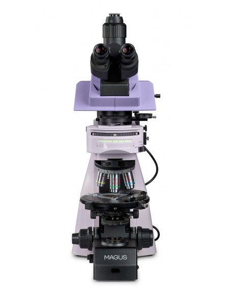
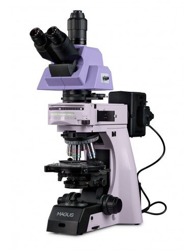
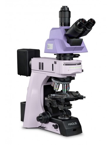
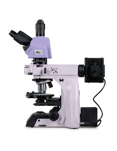
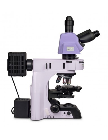
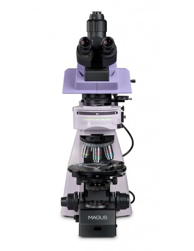
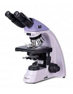
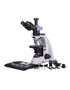
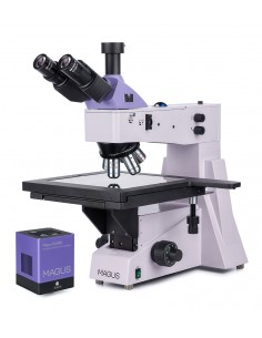
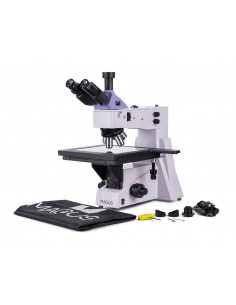
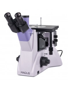
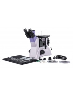
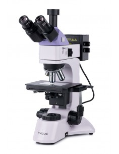
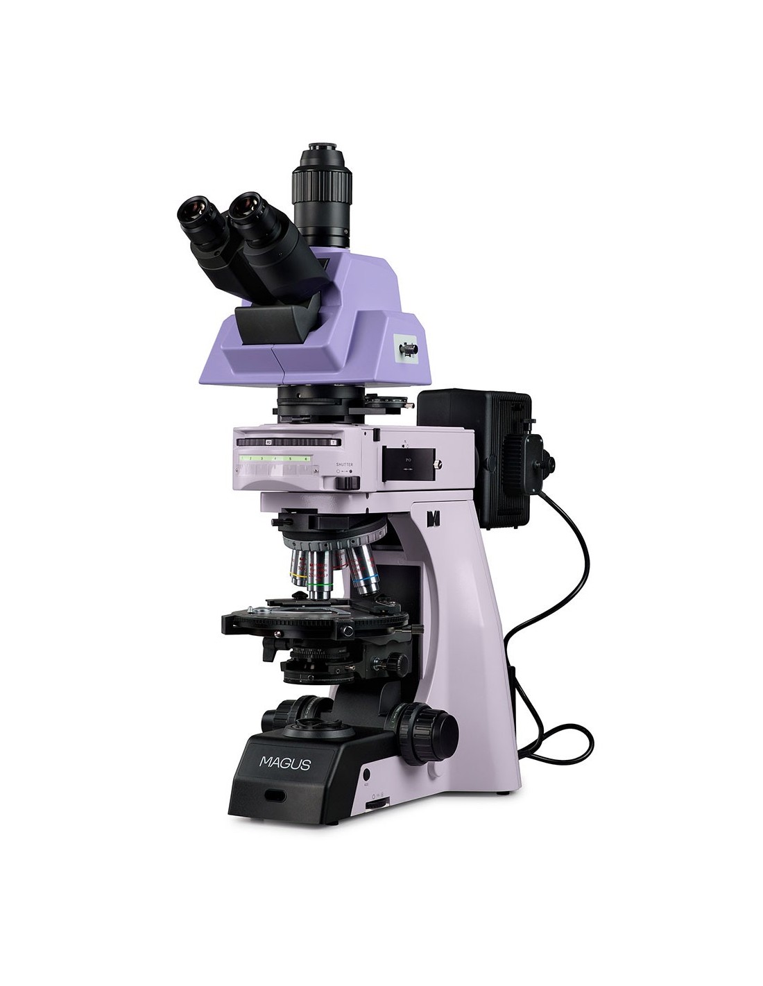
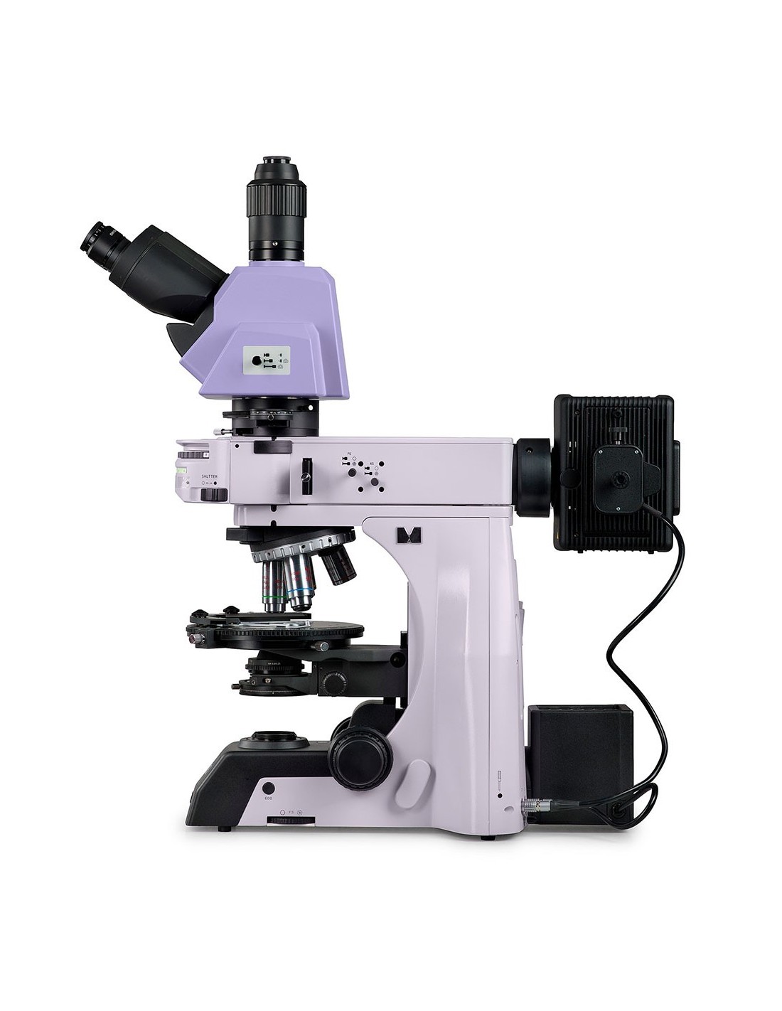
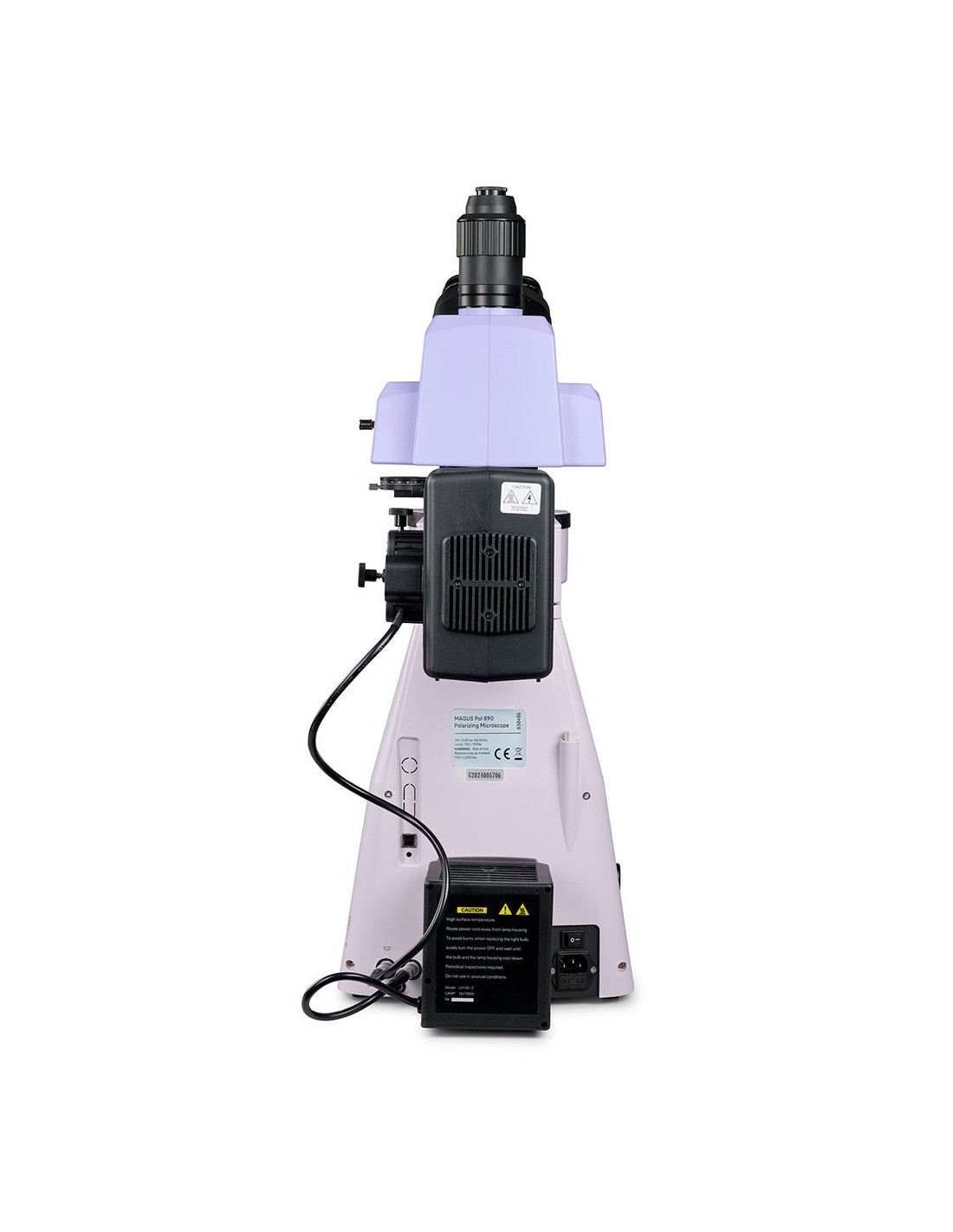
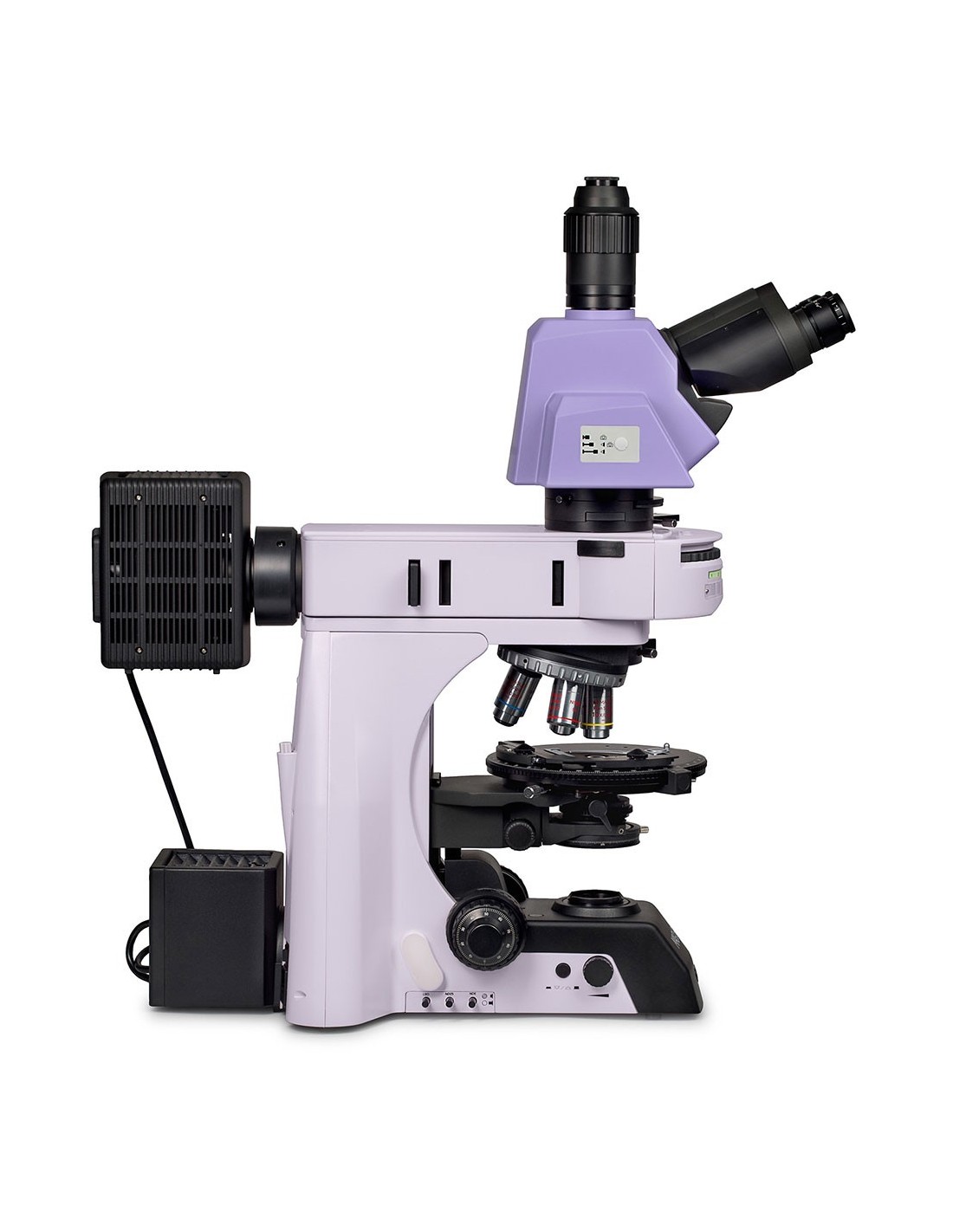
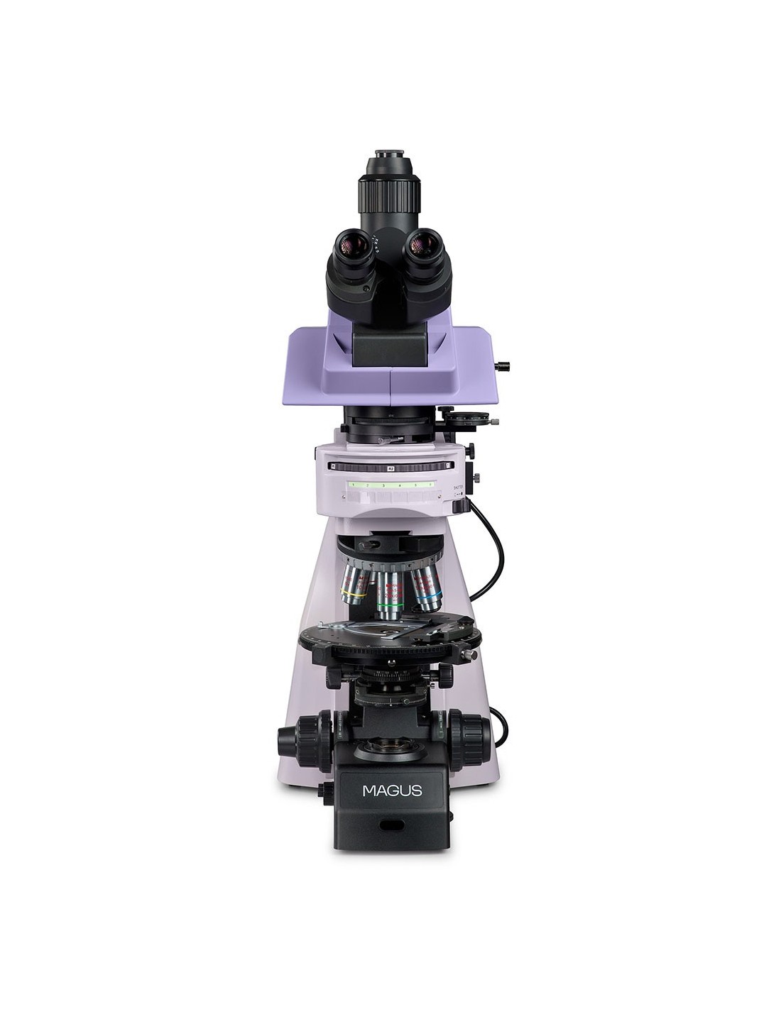
Microscope for scientific research and industrial inspection.
Designed to study objects in polarized and natural light. The installation of optional components will allow the use of dark field and transmitted light phase contrast methods as well as dark field, differential interference contrast and reflected light fluorescence.
With the transmitted light technique, it is possible to study anisotropic geological and biological samples, as well as polymer samples in thin sections.
With the reflected light technique, polished sections with one side polished can be studied. The thickness of the polished sections is arbitrary, normally 5–10 mm. The microscope allows you to examine opaque objects up to 15 mm thick.
A polarizing microscope uses the birefringence of anisotropic samples to provide an image. When plane polarized light passes through an anisotropic sample, it splits into two rays and changes the polarization plane. The analyzer brings the beam oscillations into a single plane, where they interfere. The bright, high-contrast image changes color as the stage is rotated.
Semi-apochromatic planar microscope objectives are free from internal deformations. The intermediate tube houses the analyzer and Bertrand lenses and has a housing for the compensators.
The microscope is used in crystallography, petrography, mineralogy, forensic science, medicine and other scientific fields.
Microscope head
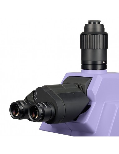
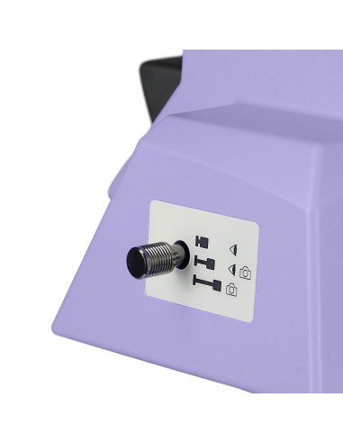
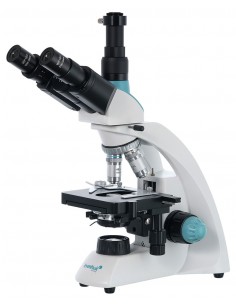
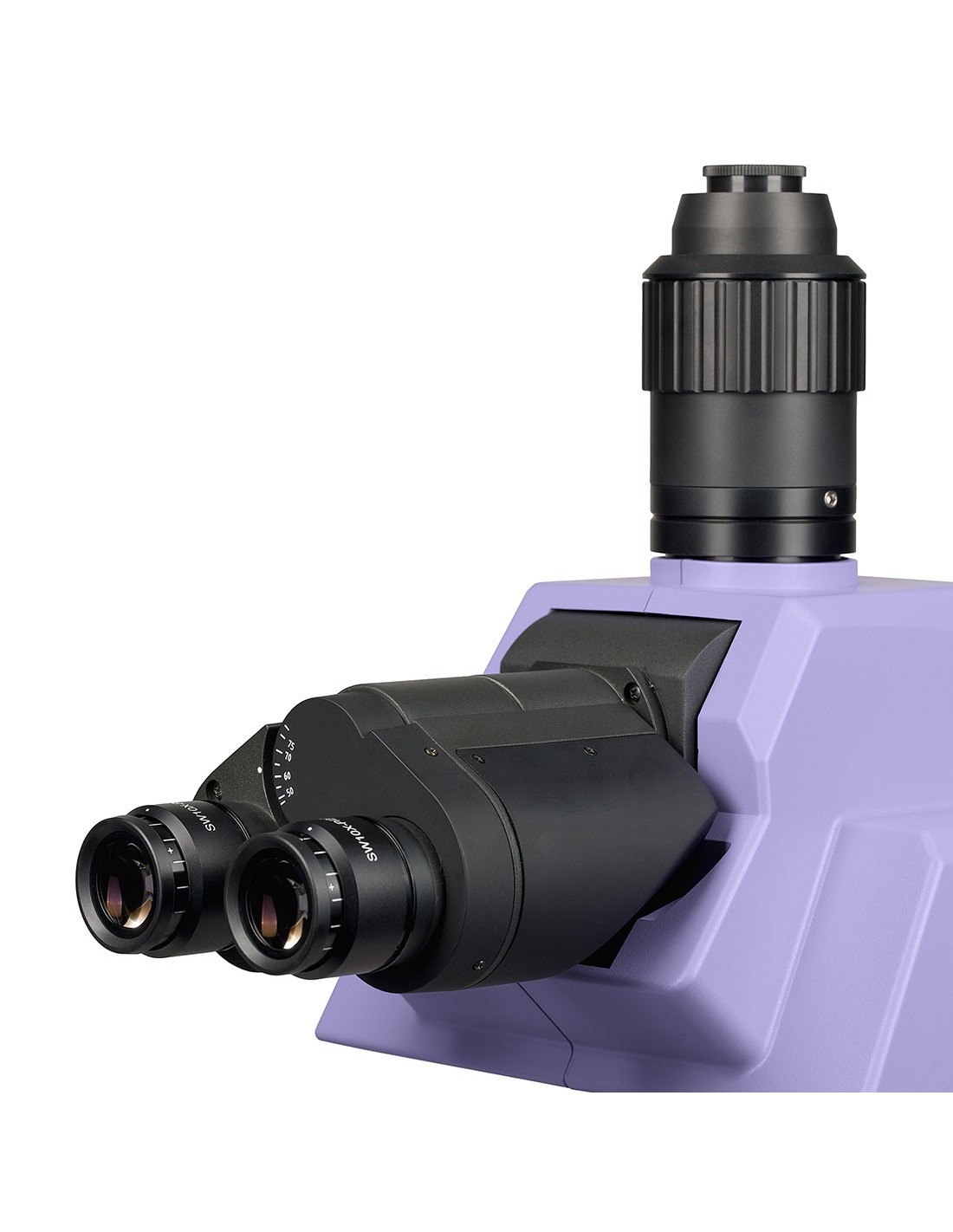
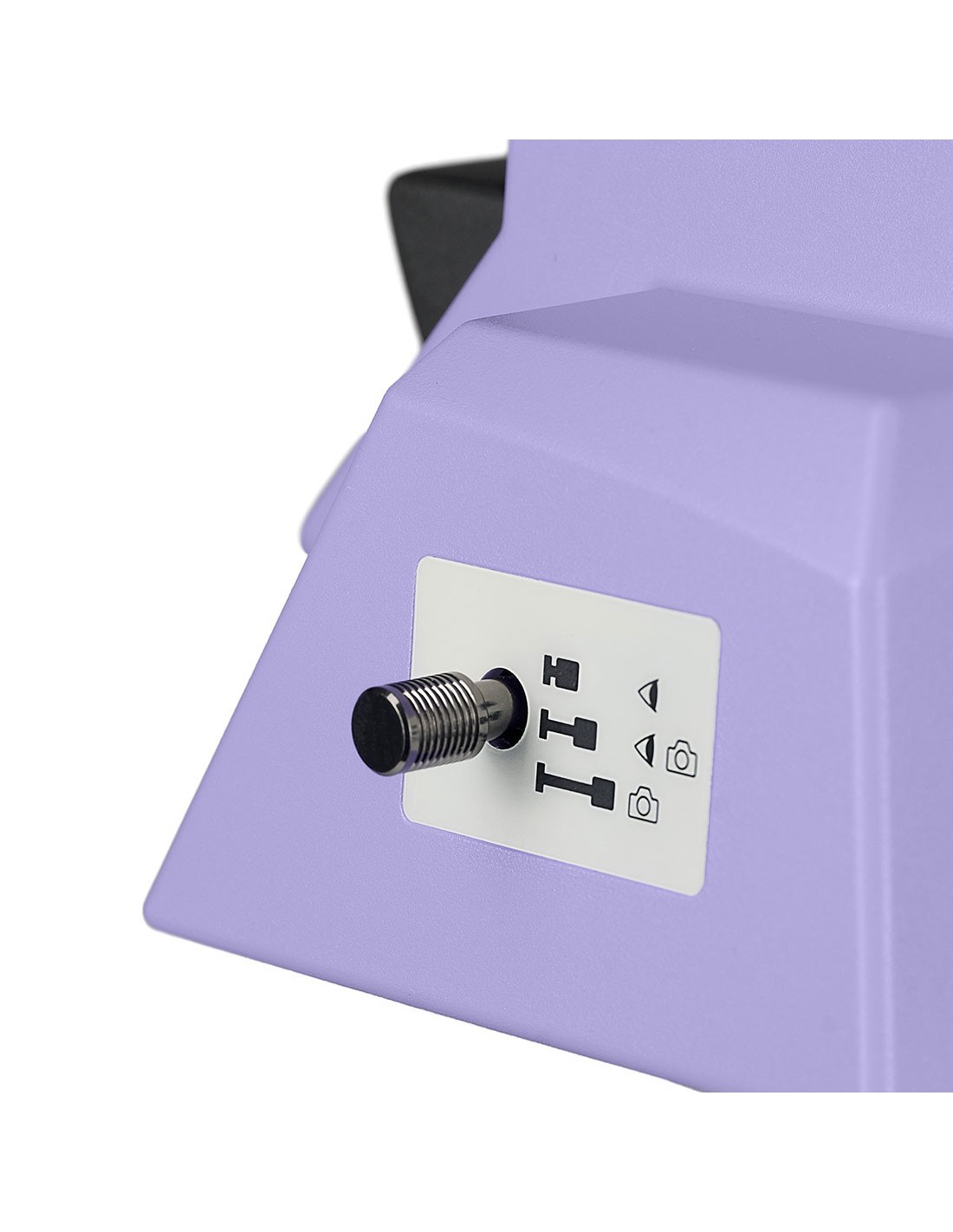
Trinocular head with infinity corrected optics. The user selects the most comfortable tilt angle of the microscope head in a range from 0° to 35°. The digital camera can be installed in the trinocular tube. The beam splits into 0/100, 100/0 or 80/20.
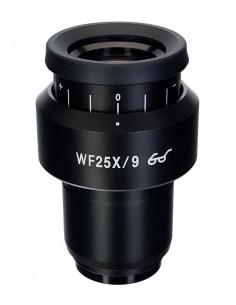
The basic kit includes 10x/22 mm eyepieces with diopter adjustment and a long eye relief for use with glasses.
Nosepiece nosepiece
Nosepiece nosepiece for five objectives. An available housing is used to centralize the light source. The user can also install an additional objective into the free nosepiece housing, which will allow him to achieve additional magnification.
The nosepiece nosepiece design (“inward”) frees up space in the front of the stage, so that the user can see the lens inserted into the optical path. The nosepiece sockets are centered to align with the optical axis of the objective and microscope.
Above the nosepiece is a socket with a plug for installing compensators.
Objectives
Planar semi-chromatic infinity objectives are specifically designed for observations in polarized light: the strain-free optics ensure that the birefringence comes from the sample and not from the optical elements. They provide better correction of chromatic and spherical aberration than plan achromatic lenses.
The 5x and 10x objectives are designed to work with or without cover glass, the 20x and 50x objectives are designed to work without cover glass. The parfocal distance is 60 mm.
Turret for installing contrast modules
Above the nosepiece there is a turret for installing contrast modules. Six modules can be installed. Rotating the turret changes the observation mode. Switching between contrast methods occurs quickly and without complex settings.
Focus mechanism
The coarse focus knobs and fine are coaxial and positioned at the bottom. The researcher can rest his hands on the table and take a comfortable position in front of the microscope.
The coarse focus lock knob helps to quickly adjust the microscope after changing the study object. The knob is located on the left side of the microscope on the same axis as the focusing mechanism.
The ring on the right side adjusts the tension of the coarse focus screw. The user adjusts the right voltage for the observations.
Stage
The stage rotates 360° to view the color of the sample changing when the polarizer and the analyzer are in a crossed orientation. The rotation angle of the coffee table can be tilted. Thanks to the vernier scale, measurements are performed with an accuracy of 0.1°.
It is possible to center the stage using two screws since the analysis of an anisotropic object in polarized light requires precise alignment of the rotation axis of the stage with the optical axis of the microscope.
Light source
100 W halogen lamps are used as illuminators for both reflected light be transmitted. Halogen lamps emit light at a color temperature that is comfortable for the eyes. The 100W lamp is bright enough to work with objectives of 4x to 100x magnification in bright field and polarized light.
Reflected light illumination
The lighting system makes the setup of Köhler lighting possible. The aperture diaphragm and field diaphragm are already centered during assembly at the factory and require no further adjustment. If necessary, the apertures can be adjusted using the centering screws. The light source is centered along three axes.
The analyzer and polarizer are used for the polarization microscopy technique. The polarizer is fixed, while the analyzer rotates 360° and has a vernier scale for precise angle setting.
A set of filters can help modify the color reproduction.
Transmitted light illumination
An adjustable field diaphragm, a centered and height-adjustable condenser with stress-free optics, an adjustable aperture diaphragm and a flip-up lens realize the adjustment of Köhler lighting. The flip-up lens is removed from the optical path for low-magnification objectives and reinserted for objectives with greater than 10x magnification. The polarizer is rotatable from 0–360°, with four rotation angles of 0°, 90°, 180°, 270° marked on the scale. The analyzer rotates 360°, the vernier scale on the analyzer guarantees the accuracy of the angle.
Köhler illumination in reflected and transmitted light
L Köhler illumination improves the image quality of the sample. With such lighting, maximum resolution can be achieved on each lens. Illumination of the field of view without obscuration from the edges is also possible. The object of study is clear and does not present image artefacts.
Study of samples in polarized light
The analyzer is inserted in the optical path to observe objects in polarized light. As the polarizer and analyzer rotate, the polarization angle changes. When you rotate the microscope stage, the refraction of light by the specimen changes depending on the polarization angle.
Bertrand lenses are used for Conopic studies.
Compensators are designed to improve the contrast of samples with weak birefringence.
Accessories
There is a line of accessories designed for this microscope.
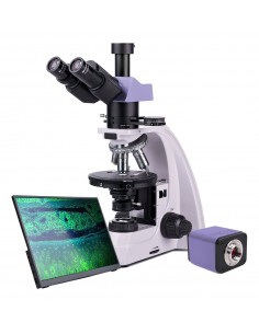
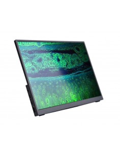
A digital camera reproduces the image of the microscope on a monitor and stores the files, while the software measures the samples in real time.
You can use a calibration slide to measure objects and combine it with an eyepiece with a scale or with camera software .
Key Features:
- Studies of opaque and transparent anisotropic samples in natural and polarized light
- Light illuminators transmitted and reflected with powerful 100 W halogen lamp providing intense natural light
- Orthoscopic observations, Bertrand lens for conopic observations, compensators, Köhler illumination in transmitted and reflected light
- Trinocular head with vertical tube for installing a digital camera and changing the tilt angle; 0/100, 100/0 or 80/20 light beam splitting
- Semi-apochromatic planar objectives without internal stresses do not introduce false optical effects into the image
- Multifunctional turret with 6 slots for installing modules of different contrast methods
- Convenient alignment of the nosepiece and stage slots
- The rotatable round stage with vernier scale for precise angle measurement of rotation, it can be fixed in a selected position
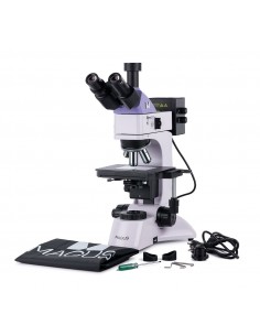
The package contains:
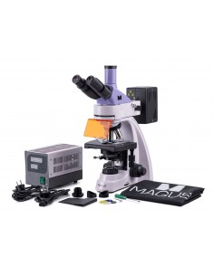
- Support with integrated power supply, setting mechanism focus, stage, condenser mount and a nosepiece revolver
- Condenser with folding lens
- Reflected light illuminator – attachment with turret and halogen lamp holder
- Transmitted light illuminator – halogen lamp holder
- Trinocular head
- Intermediate tube with Bertrand lens and analyzer
- Compensators: λ compensator; λ/4 compensator; quartz wedge
- Planar semi-apochromatic objective at infinity: 5x/0.15, parfocal height 60 mm
- Planar semi-apochromatic objective at infinity: 10x/0.30 , parfocal height 60 mm
- Planar semi-apochromatic objective at infinity: 20x/0.45, parfocal height 60 mm
- Planar semi-apochromatic objective at infinity: 50x/0 ,80, parfocal height 60 mm
- 10x/22 mm eyepiece with long eye relief and diopter adjustment (2 pcs.)
- Eyecup (2 pcs.)
- C-mount adapter
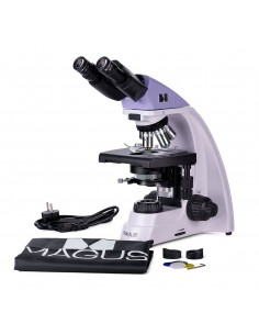
- Power cable
- Dust cover
- User manual and warranty card
Available on request:
- Digital camera
- Calibration glass
| Brand | MAGUS |
| Warranty | 5 лет |
| EAN | 5905555019529 |
| Package size (LxWxH) | 72x76x53 cm |
| Shipping weight | 31 kg |
| Type | biological, light/optical |
| Head | trinocular |
| Vertical tube | Siedentopf, beam split 0/100, 100/0, 80/ 20 |
| Tilting angle of the head | 0–35°, adjustable |
| Magnifying range | 200x to 800x |
| Eyepiece tube diameter, mm | 30 |
| 10x/22 mm, long eye relief (2 pcs.) |
| Nosepiece revolver | for 5 objectives, centering |
| Interpupillary distance, mm | 47 — 78 |
| Stage, mm | Ø190 |
| Range of movement of the stage, mm | 30/30 |
| Features stage | 360° rotatable, centering, fixable, vernier scale for measuring angles with an accuracy of 0.1°, elevated specimen holder, 1° rotation angle graduation |
| Eyepiece diopter adjustment, diopters | ±5D on each eyepiece |
| Eyepiece diopter adjustment |
| Condenser | centered and height-adjustable condenser NA 0.9/0.25 with adjustable aperture diaphragm and folding lens; stress-free optics |
| Diaphragm | adjustable aperture diaphragm, field diaphragm with adjustable iris |
| Lighting | halogen |
| Brightness adjustment | yes |
| Power | 100–240 V, 50/60 Hz, AC mains |
| Type of light source | reflected and transmitted light: lamp halogen 12 V/100 W |
| Operating temperature range, °C | 10 — 35 |
| User level | expert users, professionals |
| Assembly and installation level of difficulty | complicated |
| Polarizer | transmitted light: with 0°, 90°, 180°, 270° marked on the scale; 360° rotatable, reflected light: polarization module in the turret |
| Intermediate tube | analyzer with rotation and scale of 0–360°, Bertrand lens , housing for the installation of compensators, alternation of cognitivist/orthoscopic observations |
| Compensator | compensator λ; λ/4 compensator; quartz wedge |