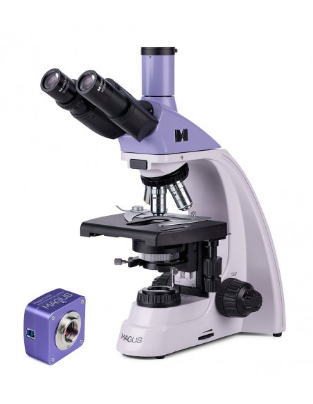
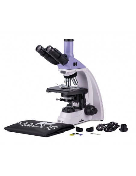
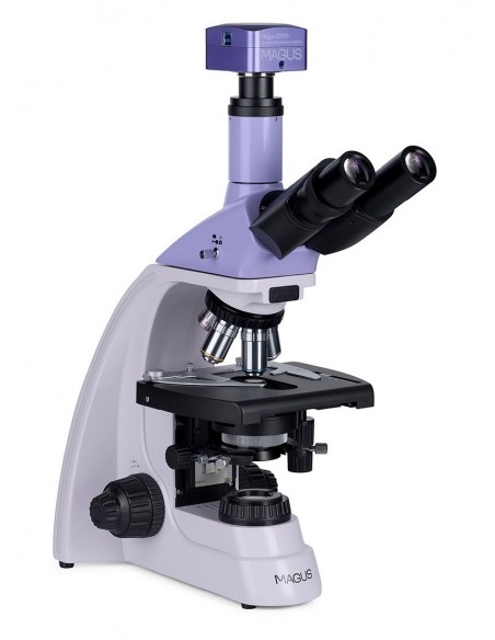
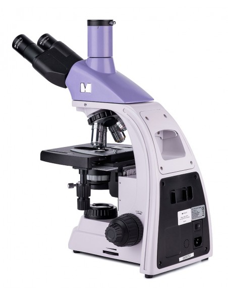
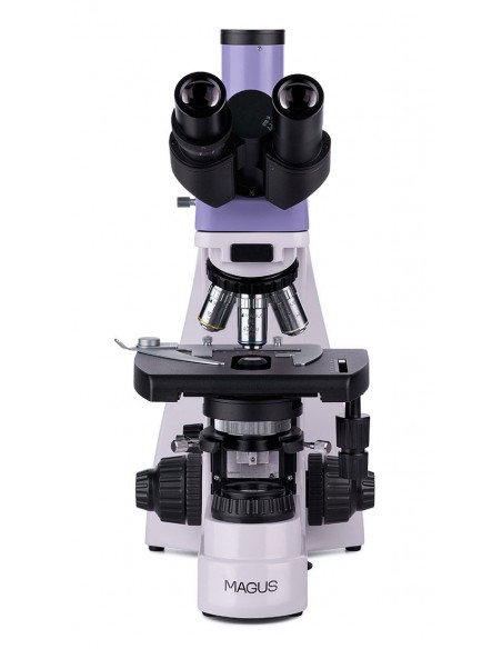
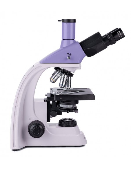
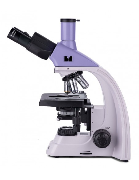
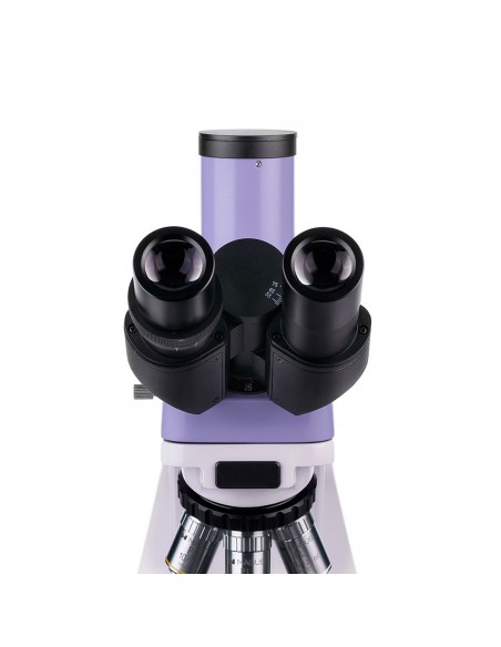
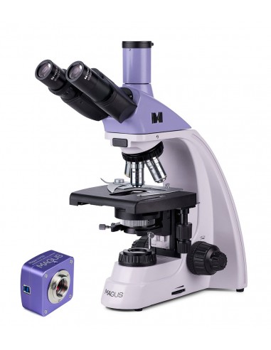
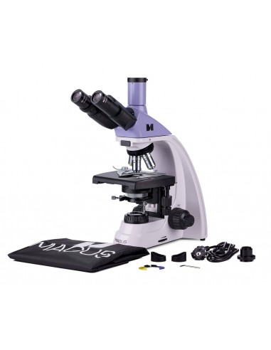
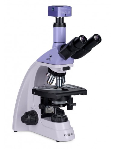
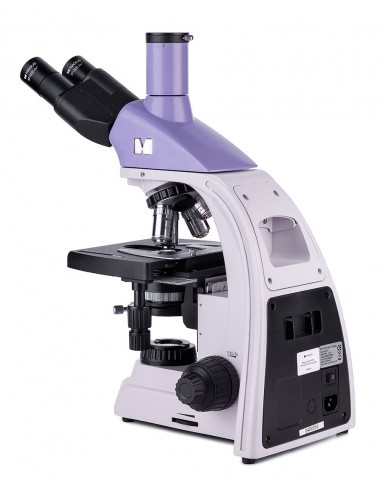
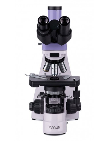
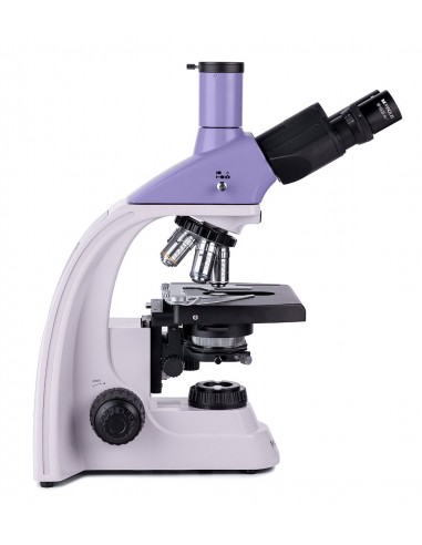
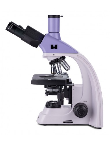
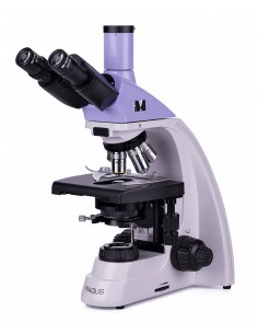
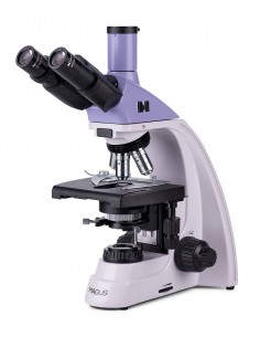
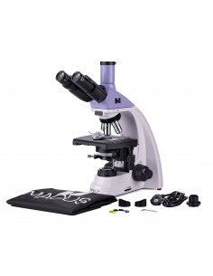
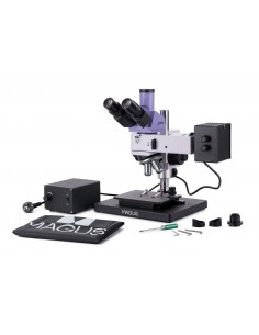
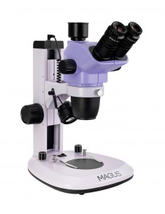
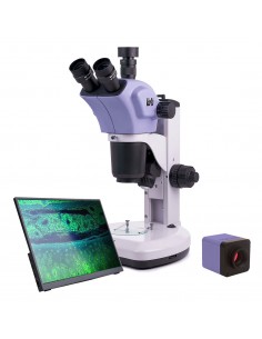
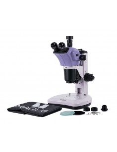
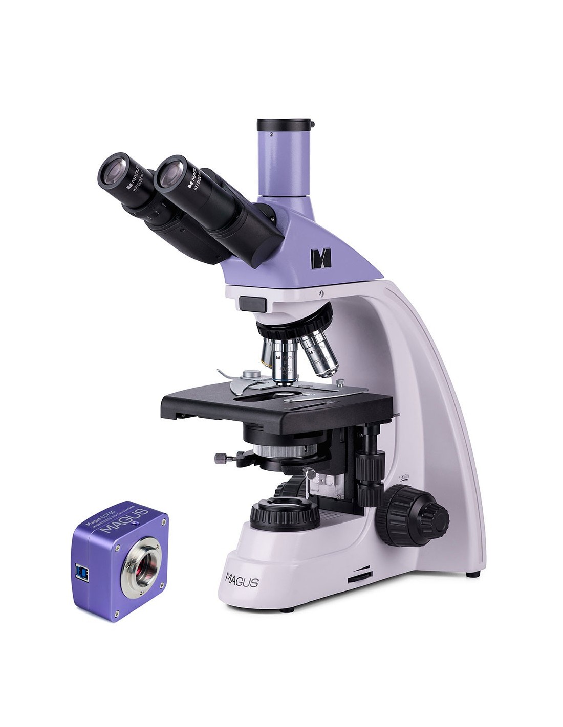
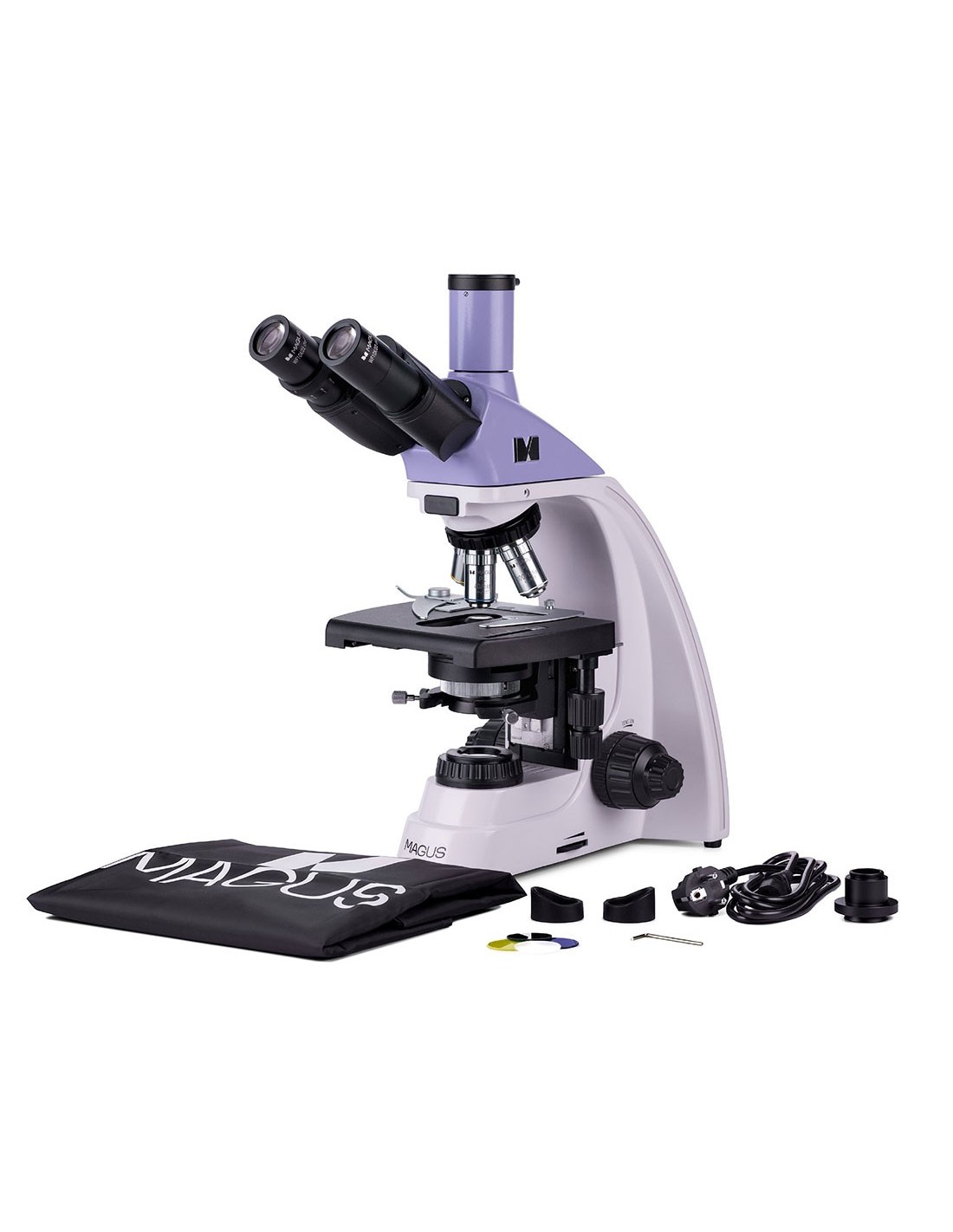
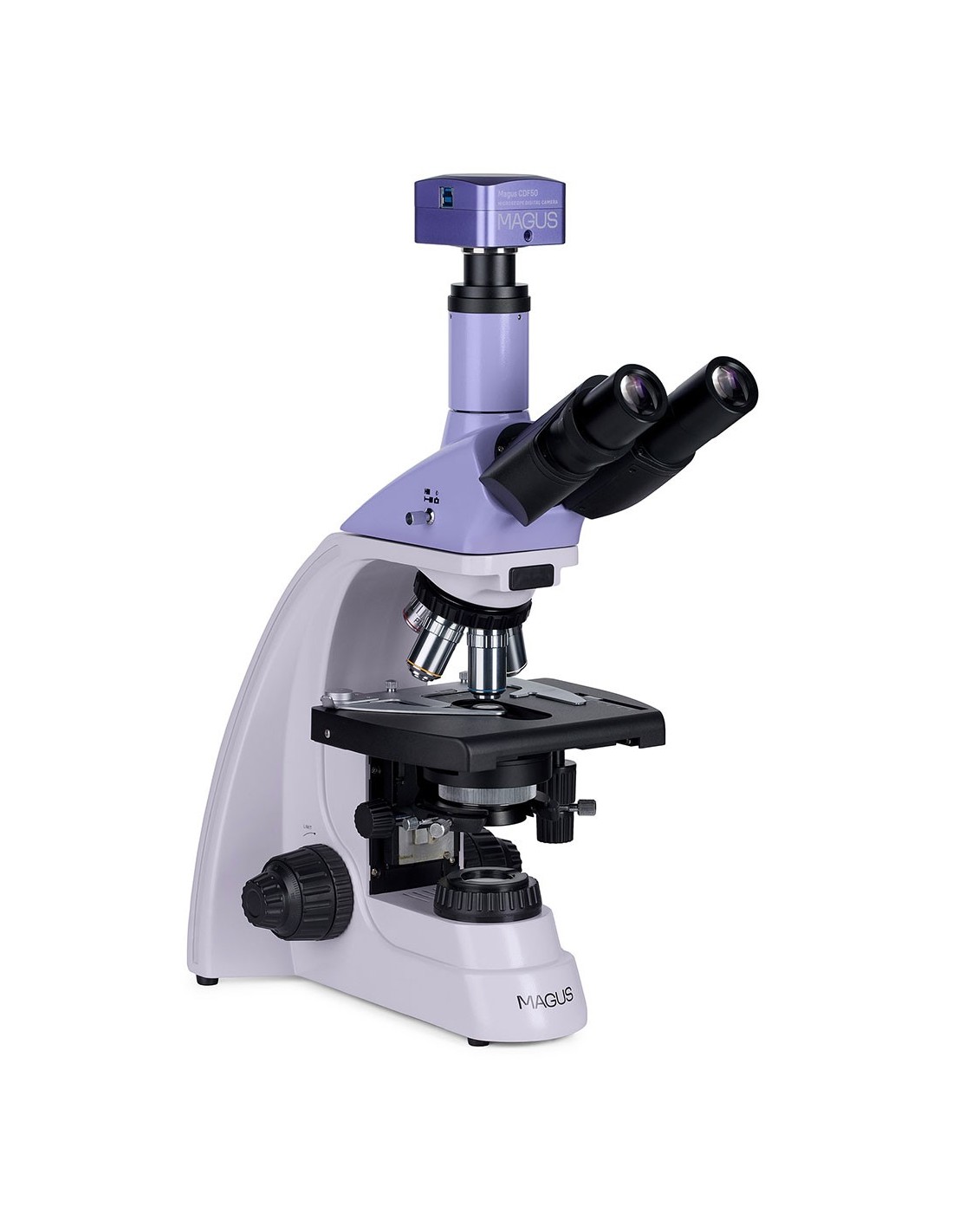
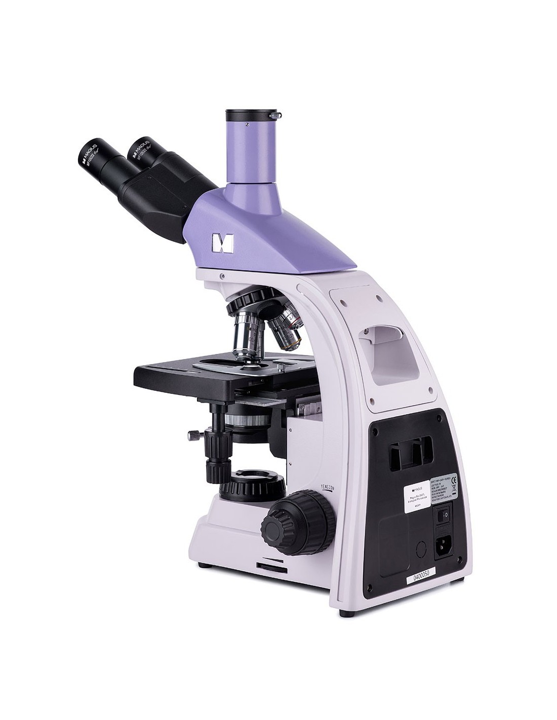
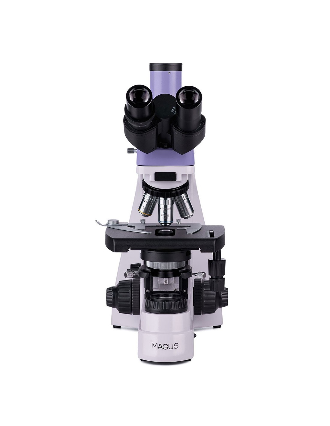
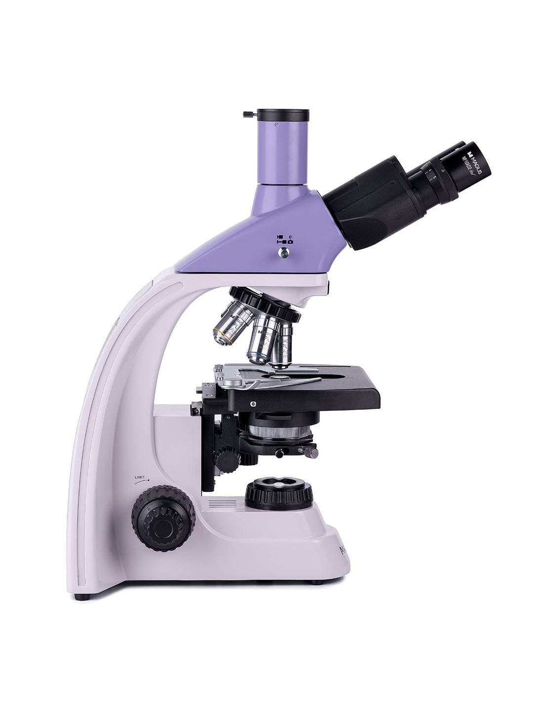
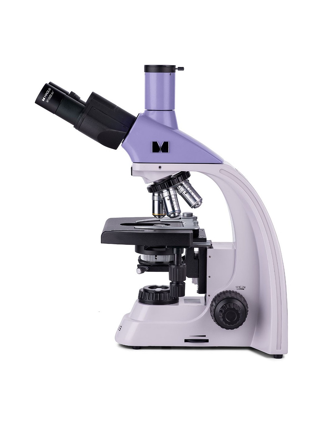
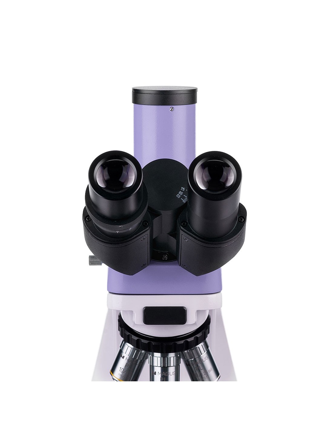
The MAGUS Bio 250TL microscope is used to examine specimens in thin sections and smears of biological materials. The observations are carried out with bright field and transmitted light microscopy techniques. A 3-watt LED light provides illumination, and the optics feature infinity-corrected plan achromatic objectives. This microscope provides the possibility of using additional accessories, including devices for observations in dark field, with polarized light or phase contrast. This microscope model is ideal for laboratory analysis, scientific research and teaching.
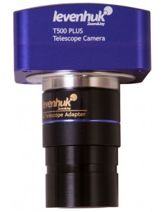
Digital camera
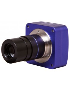
Full color camera with sensor CMOS SONY Exmor 2.1 MP digital. The sensor is produced using backlight technology to reduce light diffusion and increase the sensitivity of the matrix (8935 mV at 1/30 s). The camera produces sharp, bright images even in low-light conditions. Designed for bright- and dark-field observations using objectives with 40x, 60x and 100x magnification.
You can mount the camera on a microscope in two different ways: on the trinocular tube via a C-mount adapter or in place of the 'eyepiece on the tube via an adapter and adapter rings. We recommend using a 0.6x to 0.9x magnification adapter with your camera, which will offer the widest possible field of view without distortion. After installation, the camera connects to a computer via a USB 3.0 port with a data transfer speed of 5 Gbps.
Optics
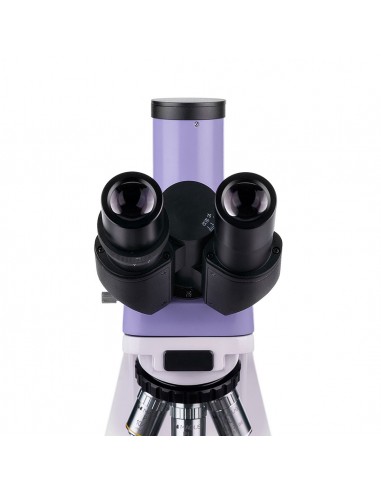
The trinocular head is composed of two optical tubes for direct observations and a vertical tube for housing a digital camera (not included). The optical tubes that house the eyepieces are inclined at 30°, for greater comfort in case of prolonged observations, and can be rotated 360° to adapt to the height of the observer. There is a diopter adjustment ring, located on the left optical tube.
The objectives are inserted in a 5-slot nosepiece revolver: 4 objectives are already mounted on the instrument, while one slot of the nosepiece remains free for 'adding an extra lens. In the free slot it is possible to insert any optional objective, allowing you to add a further magnification factor within the range of available magnifications. The user can see which objective is inserted in the optical path during observation, since the nosepiece is oriented towards the inside of the instrument. Furthermore, this configuration keeps the space above the stage free.
The magnification range of the microscope is from 40x to 1000x (only in the basic configuration). By adding extra objectives, the higher magnification factor will be increased.
Illumination
LEDs mounted inside the microscope base provide intense and uniform illumination of the observed object. The color temperature remains unchanged even when the brightness varies. The lighting has a power of 3 W. The useful life cycle of the LEDs reaches 50,000 hours. The illumination system is powered by AC mains electricity.
By adjusting the position of the Abbe condenser and opening or closing the aperture diaphragm and the field diaphragm, the Köhler illumination can be configured. It is also possible to use sliders for dark field or phase contrast observation techniques (the condenser is equipped with a dedicated slot). Thanks to the use of the sliders, it will be very quick to switch from one observation method to another.
Stage and focusing mechanism
The stage It does not have a positioning rack. As a result, there is no cumbersome mechanism to disturb observations and the microscope takes up less space on the workbench. The mechanical insert that moves the sample is removable (useful in case of manual scans).
The coarse and fine focusing mechanisms are used to adjust the sharpness. The relevant knobs are located on both sides of the microscope. Given their position close to the base, it will not be necessary to keep your arms in mid-air to adjust the sharpness. Your hands will be able to remain comfortably resting on the table. The coarse focus is equipped with a fixing knob and the tension of the coarse screw can be adjusted. Sharpness adjustment is fluid and effortless.
Accessories
The potential of the MAGUS Bio 250TL microscope can be increased thanks to the line of dedicated accessories, which includes eyepieces, objectives, calibration slides, digital cameras and instruments for observation in dark field, with polarized light or phase contrast.
Key features of the microscope:
- Trinocular head, nosepiece nosepiece for 5 objectives, infinity corrected planachromatic optics
- The optical tubes of the eyepieces rotate 360° to easily adapt to the observer's height
- li>
- Left optical tube with diopter adjustment ring; coarse and fine focus (coarse and fine screw)
- The coarse focus is equipped with a fixing knob and the tension of the coarse screw can be adjusted
- The capacitor Abbe is centerable and height-adjustable, and the presence of an aperture diaphragm and a field diaphragm allow you to configure the Köhler illumination
- Light source for transmitted light observations: 3 W brightness LED adjustable, powered by 220 V AC mains electricity
- Possibility to install additional accessories
Key features of the camera:
- 2.1 inch back-illuminated CMOS sensor, high matrix sensitivity
- Suitable for dark or bright field observations with 40x, 60x and 100x objectives
- Installed on the trinocular tube or on the optical tube of a microscope
- Full HD resolution 1920x1080 px with a frame rate of 96 fps, perfect for studying moving objects
- Connection to PC via USB 3.0 port, data transfer speed of 5 Gbps
- Software and drivers included
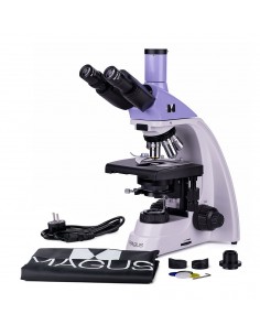
The package contains:
- MAGUS CDF50 digital camera (digital camera, USB cable, installation CD with drivers and software, user manual and warranty card)
- Base with power input, light source for transmitted light observations , focusing mechanism, stage, condenser attachment and nosepiece
- Abbe condenser
- Trinocular head
- Infinity planachromatic objective: 4x/0, 10
- Infinity plan achromatic objective: 10x/0.25
- Infinity plan achromatic objective: 40x/0.65 (with spring mechanism)
- Infinity planachromatic objective: 100x/1.25 oil (with spring mechanism)
- 10x/22 mm eyepiece with long eye relief (2 pcs.)
- Eyecup (2 pcs.)
- Filter (4 pcs.)
- C-mount adapter
- Vial of immersion oil
- Power cord for AC mains
- Dust cover
- User manual and warranty card
Available on request:
- 10x/22 mm eyepiece with graduated scale
- 12.5x/14 mm eyepiece (2 pcs.)
- 15x/15 mm eyepiece (2 pcs.)
- Eyepiece 20x/12 mm (2 pcs.)
- Eyepiece 25x/9 mm (2 pcs.)
- Planachromatic infinity objective: 20x/0 ,40
- Planachromatic infinity objective: 60x/0.80 (with spring mechanism)
- Phase contrast device
- Phase contrast slider
- Dark field capacitor with NA 0.9
- Oil dark field capacitor with NA 1.36–1.25
- Dark field slider
- Polarization Tools
- Calibration Glass
- LCD Monitor
| Brand | MAGUS |
| Warranty | 5 лет |
| EAN | 5905555018065 |
| Package size (LxWxH) | 43x27x63 cm |
| Shipping weight | 11.1 kg |
| Delimiter | MAGUS Bio 250TL biological microscope |
| == = Delimiter === | Microscope Specifications |
| Type | biological, light/optical, digital |
| Trinocular head |
| Head inclination angle | 30° |
| Magnification, x | 40–1000 in the basic configuration (*optional: 40–1200/1250/1500/1600/2000/2500) |
| Magnifying range |
| Eyepiece tube diameter, mm | 30 |
| Eyepieces | 10х/22 mm, eye relief: 10 mm (*optional: 10x/22 mm with graduated scale, 12.5x/14; 15x/15; 20x/12; 25x/9) |
| Infinity planachromatic | objectives: 4x/0.10; 10x/0.25; 40xs/0.65; 100xs/1.25 (oil); parfocal distance: 45 mm (*optional: 20x/0.40; 60хs/0.80) |
| Nosepiece nosepiece | for 5 objectives |
| Working distance, mm | 21 (4x); 5 (10x); 0.66 (40xs); 0.36 (100xs); 8.8 (20x); 0.465 (60xs) |
| Interpupillary distance, mm | 48 — 75 |
| Stage, mm | 180x150 |
| Range of movement of the table, mm | 75/50 |
| two-axis mechanical translator, without positioning rack |
| Eyepiece diopter adjustment, diopters | ± 5 (on left barrel) |
| Condenser | Abbe condenser, NA 1.25, adjustable centering, adjustable height, adjustable aperture diaphragm, with slot for dark field slider and phase contrast slider, dovetail mount |
| Diaphragm | adjustable aperture diaphragm, field diaphragm with adjustable iris |
| Focus | coaxial, coarse focus (21 mm, 39.8 mm/turn, with fixing knob and tension adjustment knob micrometer screw) and fine focus (0.002 mm micrometer screw) |
| Illumination | LED |
| yes |
| Power supply | 220±22 V, 50 Hz, AC mains |
| Type of light source | 3 W LED |
| Optical filters | yes |
| Operating temperature range, °C | 5 — 35 |
| Possibility to connect additional equipment |
| td> | phase contrast devices (condenser and objectives), dark field condenser (dry or oil immersion), polarization instruments (polarizer and analyzer) |
| expert users, professionals |
| Assembly and installation level of difficulty | complicated |
| Application | laboratory/medical |
| Lighting position | lower |
| Search method | bright field |
| Set with pencil case/case/bag | dust cover |
| Weight, kg | 8 |
| Dimensions, mm | 200x436x400 |
| === Delimiter === | Digital camera |
| Sensor | SONY Exmor CMOS |
| Color/monochrome | color |
| Megapixel | 2.1 |
| Maximum resolution, px | 1920x1080 |
| Sensor size | 1/1.2'' (11.14x6.26 mm) |
| Pixel size, µm | 5.8x5.8 |
| Light sensitivity | 8935mV with 1/30s |
| Signal to noise ratio | 0.6mV with 1/30s |
| Exposure time | 0.014 ms – 15 s |
| Video recording | yes |
| Frame rate, fps@resolution | 96@1920x1080 |
| Installation location | trinocular tube, optical tube instead of eyepiece |
| Image format | *.jpg, *.bmp, *.png, *.tif |
| Video format | recording: *.wmv, *.avi, *.h264 (Windows 8 and later), *h265 (Windows 10 and later) |
| Spectral range, nm | 380-650 (integrated IR filter) |
| Shutter type | ERS (electronic curtain shutter) |
| White balance | manual, automatic |
| Exposure control | manual, automatic |
| Software features |
| Software | MAGUS View |
| Output | USB 3.0, 5 Gb/s |
| System requirements | Windows 8/10/11 (32 bit and 64 bit ), Mac OS | Mount type | C-pitch |
| Body | CNC aluminum alloy |
| Camera power supply | DC, 5 V, from computer USB port; 12 V, 3 A adapter for the Peltier cell |
| Camera operating temperature range, °C | -10 — 50 |
| Humidity operating range, % | 30 — 80 |