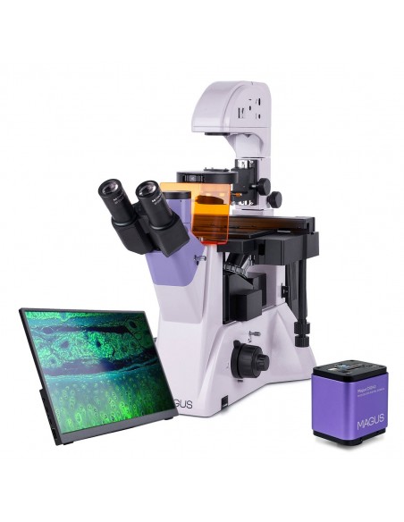
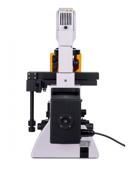
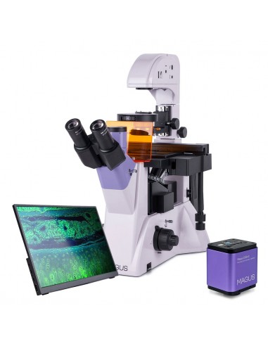
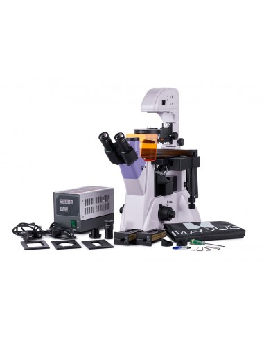
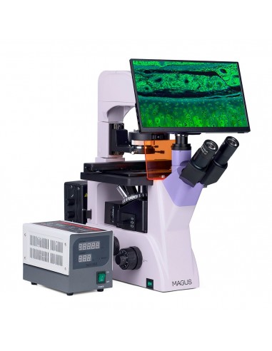
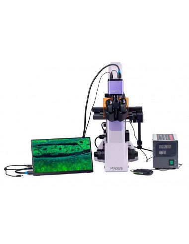
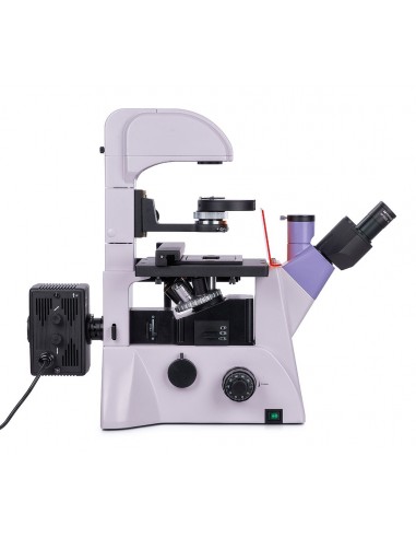
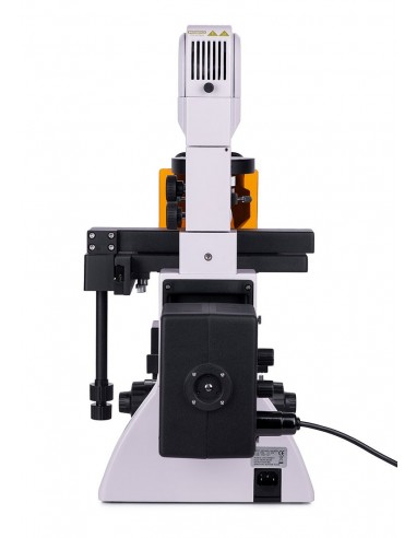
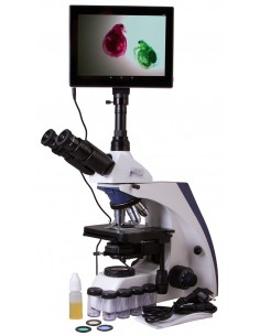
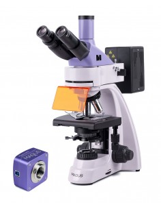
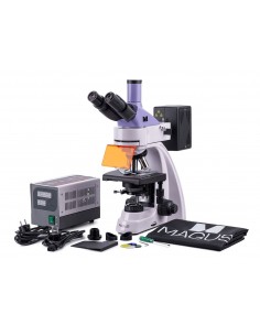
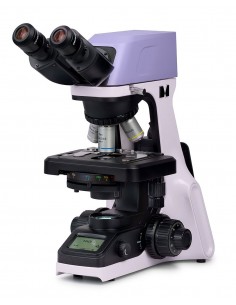
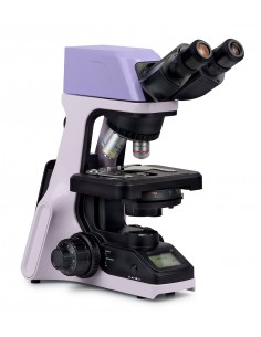
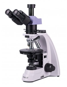
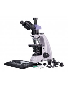
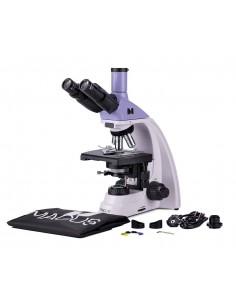
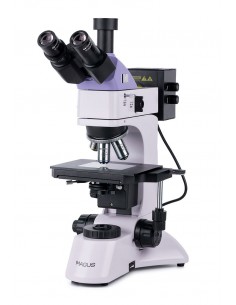
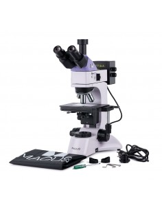
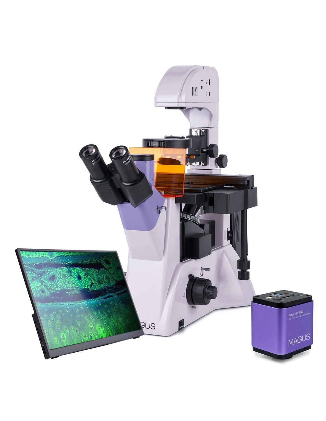
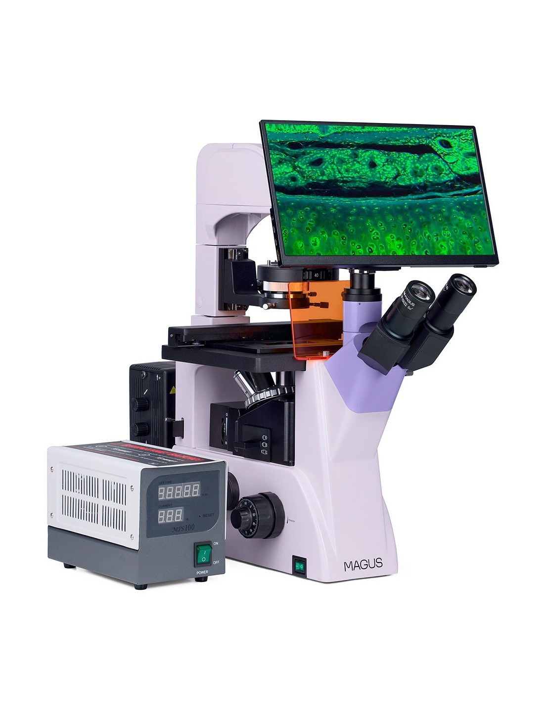
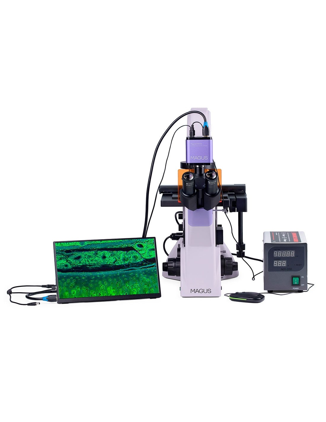
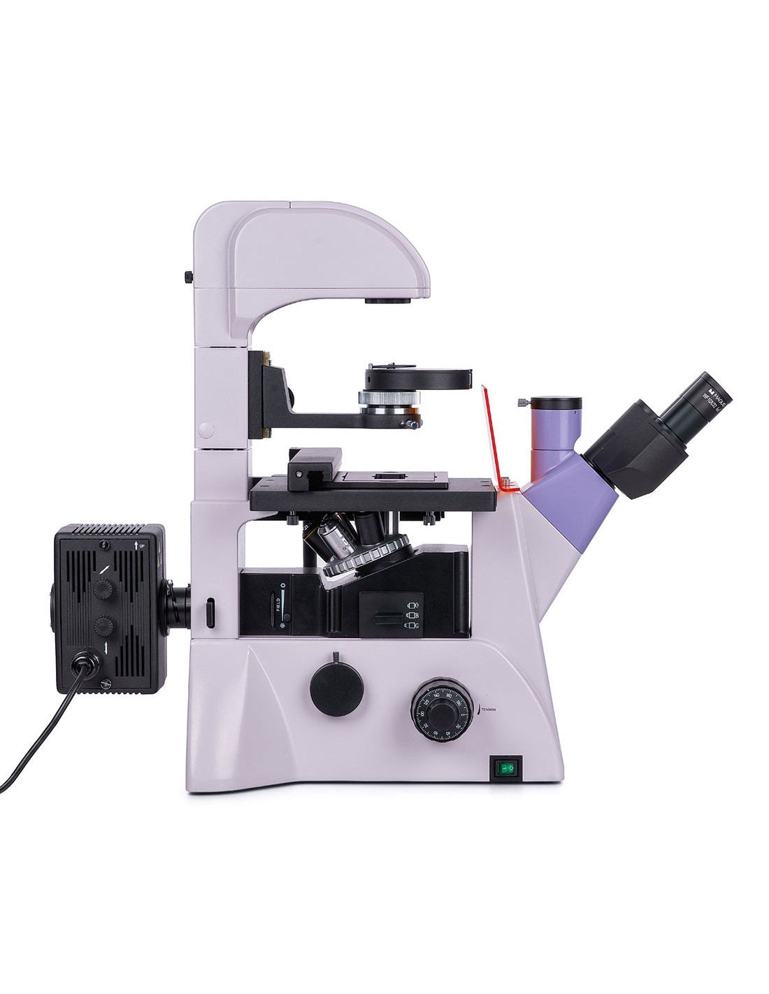
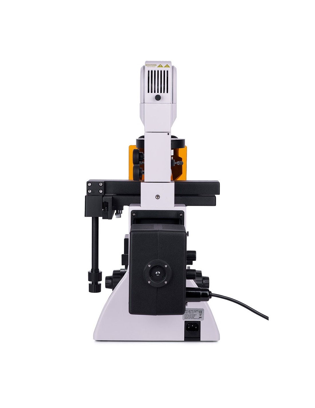
The MAGUS Lum V500 inverted fluorescence microscope is designed for observing samples in laboratory glassware with a height of up to 55 mm and a bottom thickness of up to 1.2 mm. By tilting the illuminator support, it becomes possible to use larger laboratory glassware, up to 165 mm. Samples can be observed in reflected light (fluorescence microscopy technique) and in transmitted light (with bright field and phase contrast microscopy). The main fields of application of this microscope are: medicine, pharmacology, biochemistry, epidemiology, etc.
Digital camera
MAGUS CHD40 is an HDMI digital camera with three video interfaces and automatic switching between Full HD and 4K based on monitor resolution.
The camera is equipped with an 8 MP sensor and produces realistic images at 4K resolution (3840x2160 px) when connected via HDMI or USB 3.0. When connected via Wi-Fi, the image quality is Full HD (1920x1080 px).
The camera uses an HDMI interface to connect directly to a TV, monitor or projector. In this way, the camera operates independently without a connection to a PC. The HDMI interface enables fast and stable speed transfer from the camera to the external screen. You can connect the camera to a PC via Wi-Fi or USB 3.0. Video is recorded at 30 fps.
The camera combines high FPS rate and high HDMI bandwidth. The videos therefore appear brilliant without freezing or skipping between frames. At maximum resolution, the image is very detailed, moving objects are visible without errors, and the moving object is displayed without delays.
Monitor
The MAGUS MCD40 monitor is designed to use a MAGUS microscope display system. It is connected to a camera mounted on the microscope to display real-time images. Supports cameras with HDMI connection and 4K resolution. The screen is 13.3 inches. The IPS matrix offers bright images with wide viewing angles. If you look at the display from the side the color reproduction is not distorted. You can place the display on a table or shelf via a foldable stand or install it directly on the camera or microscope base.
Optics
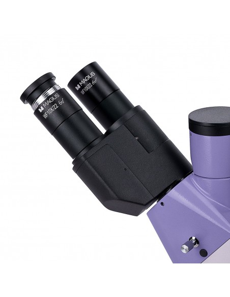
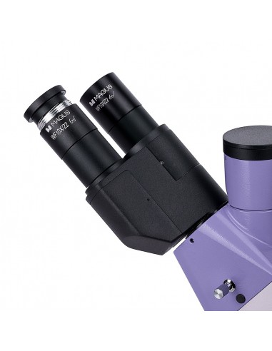
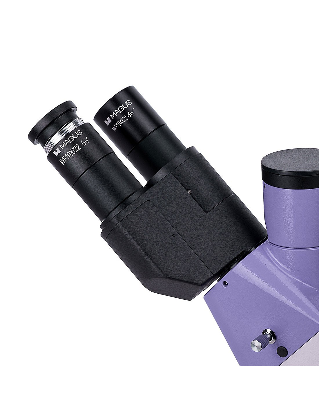
This microscope is equipped with a trinocular head with vertical optical tube. There is a side camera port on the microscope frame. This design choice allows you to simultaneously install a digital camera and a monitor (not included) on the microscope. The beam splitter of the trinocular head has a ratio of 80/20, while that of the microscope frame is 100/0 and 0/100. The eyepiece tubes are rotatable through 180°, which allows the eye relief to be adjusted to suit the user.
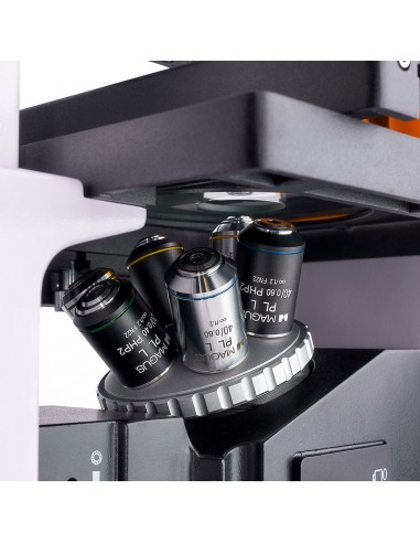
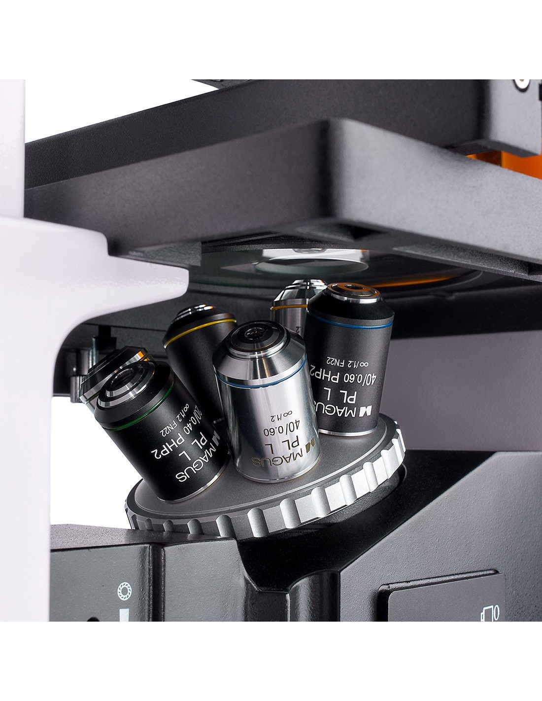
The nosepiece is placed on a stand underneath the stage. It is equipped with six free slots, for housing as many lenses. The package includes three objectives for phase contrast and three objectives for fluorescence and brightfield observations. These are planachromatic objectives with a long working distance, designed for laboratory glassware with a 1.2 mm thick bottom.
Illumination
The source of Reflected light is a 100W mercury vapor lamp. It is placed in a lamp holder for better heat dissipation and less risk of overheating during extended use. This lamp emits light in a wide range of wavelengths with various peak intensities, making it perfect for working with a variety of fluorophores. Fluorescence filters: ultraviolet (UV), violet (V), blue (B), and green (G). The mercury vapor lamp illuminates the sample intensely, providing good visibility and can be easily replaced when necessary.
The transmitted light is provided by a 30 W halogen lamp. The illumination provided is intense and with a warm color temperature. As a result, observation through each of the lenses is always efficient and comfortable for the eyes. The microscope has a phase contrast condenser with four slots: one is used for brightfield observations and the other three are designed for 10x, 20x and 40x phase contrast objectives. Phase contrast rings can be centered. To switch from bright field observation to phase contrast techniques, and vice versa, simply rotate the disk. This is particularly practical for professional researchers, as it saves time.
Application of Köhler illumination for transmitted and reflected light observations. Using this method allows you to obtain clear and sharp images, in high resolution, without artifacts or darkening of the edges.
Focusing stage and mechanism
The stage is fixed and is equipped with a mechanism for moving the laboratory glassware on which the sample is positioned. Laboratory glassware can be moved along the horizontal plane, along two axes perpendicular to each other. The movement is smooth and precise and the mechanism includes four holders for accommodating glassware of various sizes. The sharpness of the microscope image is adjusted by rotating the coarse and fine focus knobs (macro and fine screw). The coarse focus is equipped with a fixing knob and the tension of the coarse screw can be adjusted. The focus knobs are coaxial and are located on both sides of the frame.
Accessories
This product line includes additional accessories for the microscope MAGUS Lum V500: eyepieces, digital cameras and calibration slides. Fluorescence microscopy in reflected light and bright field and phase contrast microscopy in transmitted light
Key features of the microscope:
- Possibility of observing samples inside laboratory glassware with a height of up to 55 mm, or 165 mm with inclined support
- 100 W mercury vapor lamp for reflected light observations, 30 W halogen lamp for transmitted light observations, configurable Köhler illumination
- Four fluorescence filters: ultraviolet (UV), violet (V), blue (B) and green ( G)
- 180° rotatable trinocular head. Two options for installing a digital camera and monitor: 80/20 beam splitter (trinocular head) or 100/0 or 0/100 beam splitter (microscope frame)
- Stage with slots for Petri dishes and laboratory glassware of various diameters
- Phase contrast condenser with bright field aperture and three phase contrast apertures
Key camera features:
- The camera works independently without a connection to a PC via HDMI interface. You can connect to a PC via Wi-Fi and USB 3.0 interface
- Automatic switching between 4K and Full HD based on monitor resolution
- 30 fps for observing objects in motion, video recording, and sample movement without lag or stuttering
- The color back-illuminated SONY Exmor/ Starvis CMOS sensor delivers images with low noise and high light sensitivity even in low light conditions. You can get sharper, brighter and more saturated color images.
- Software for recording and editing photos and videos, external display functions, linear and angular measurements
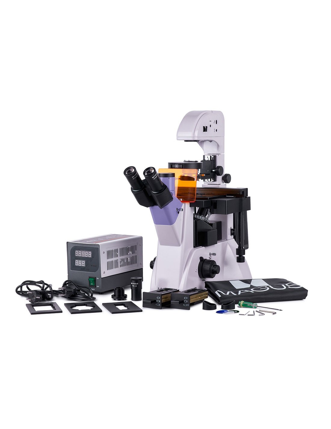
The package contains:
- MAGUS CHD40 digital camera (digital camera, HDMI cable (1.5 m), USB 3.0 cable (1.5 m), mouse with USB, 32GB SD Memory Card, Wi-Fi USB Adapter (2pcs), 12V/1A AC Power Adapter (Euro), Installation CD with Drivers and Software, User Manual and Warranty Card)
- MAGUS MCD40 LCD monitor
- Base with power input, transmitted light light source and condenser, focusing mechanism, stage and nosepiece
- Setting unit fluorescence
- Mercury vapor lamp housing
- Trinocular head
- Infinity planachromatic objective: PLL 10x/0.25 WD 4.3 mm
- Infinity planachromatic objective: PLL 10x/0.25 WD 4.3 mm
- li>
- Infinity plan achromatic objective: PLL 20х/0.40 WD 8.0 mm
- Infinity plan achromatic objective: PLL 40х/0.60 WD 3.5 mm
- Plan achromatic infinity objective: PLL 10x/0.25 PHP2 WD 4.3 mm
- Plan achromatic infinity objective: PLL 20x/0.40 PHP2 WD 8.0 mm
- Infinity planachromatic objective: PLL 40x/0.60 PHP2 WD 3.5 mm
- 10x/22 mm eyepiece with long eye relief (2 pcs.)
- Screen UV
- 1x C-mount adapter
- Allen key
- Mercury vapor lamp power supply
- Power cable
- Reflected light illuminator power cable
- Dust cover
- User manual and warranty card
Available upon request :
- 10x/22 mm eyepiece with graduated scale
- 12.5x/14 mm eyepiece (2 pcs.)
- Eyepiece 15x/15 mm (2 pcs.)
- Eyepiece 20x/12 mm (2 pcs.)
- Eyepiece 25x/9 mm (2 pcs.)
- Calibration glass
| Brand | MAGUS |
| Warranty | 5 лет |
| EAN | 5905555018225 |
| Package size (LxWxH) |
| td> | 38x61x82 cm |
| Shipping weight | 28.75 kg |
| Delimiter | MAGUS Lum V500 inverted fluorescence microscope |
| Type | biological, light/optical |
| trinocular |
| Vertical tube | Siedentopf, 180° rotatable |
| Magnification, x | 100–400 in basic configuration (*optional: 40–500/600/800/1000) |
| Tube diameter eyepiece, mm | 30 |
| Eyepieces | 10х/22 mm, eye relief: 10 mm (*optional: 10x/22 mm with graduated scale, 12.5x/14; 15x/15; 20x/12; 25x/9) |
| Objectives | plan achromatic objectives at infinity (∞): PLL 10x/0.25/4.3; PLL 20x/0.40/8.0; PLL 40x/0.60/3.5; phase contrast: PLL 10x/0.25/4.3 PHP2; PLL 20x/0.40/8.0 PHP2; PLL 40x/0.60/3.5 PHP2; parfocal distance: 45 mm |
| Nosepiece revolver | for 6 objectives |
| Working distance, mm | 4.3 (10х); 8.0 (20x); 3.5 (40x) |
| Interpupillary distance, mm | 48 — 75 |
| Stage, mm | 227x208 |
| Range of movement of the stage, mm | 77/134.5 |
| Table features | fixed, with Ø118 mm glass plate and mechanical slider; plate holder: 86x129.5, Ø90 mm; 34x77.5 mm, Ø68.5 mm; 57x82 mm, Ø60 mm; 29x77.5 mm, Ø35 mm |
| Capacitor | NA 0.6; working distance: 55 mm; phase contrast turret; with fixing screws |
| Diaphragm | adjustable aperture diaphragm, field diaphragm with adjustable iris |
| Focus | coaxial, coarse focus (with micrometer screw tension adjustment knob and fixing knob) and fine focus (0.002 mm micrometer screw) |
| Lighting | halogen, fluorescent |
| Brightness adjustment | yes |
| Power supply | AC mains, 85–265 V, 50/60 Hz |
| Type of light source | reflected light: 100 W mercury vapor lamp; transmitted light: 12 V halogen lamp, 30 W |
| Optical filters | yes |
| Operating temperature range, °C | 5 — 35 |
| Special features | phase contrast condenser (turret) with available housing and phase contrast ring plates for 10x, 20x, and 40x objectives; auxiliary centering microscope |
| User level | expert users, professionals |
| Level of difficulty assembly and installation | complicated |
| Fluorescent module | filters: ultraviolet (UV), violet (V), blue (B), green (G) |
| Fluorescence filter: filter type/excitation wavelength/dichroic mirror/emission wavelength | ultraviolet ( UV), 320–380 nm/425 nm/435 nm; violet (V) 380–415 nm/455 nm/475 nm; blue (B) 450–490 nm/505 nm/515 nm; green (G) 495–555 nm/585 nm/595 nm |
| Application | laboratory/medical |
| Illumination position | double |
| Research method | bright field, phase contrast microscope, fluorescence |
| Set with pencil case/case/bag | dust cover |
| === Delimiter ===< /td> | Digital camera |
| Sensor | Sony Exmor/Starvis CMOS |
| A color/monochrome | color |
| Megapixel | 8 |
| Resolution maximum, px | 3840x2160 |
| Sensor size | 1/1.2'' (11.14x6.26 mm)< /td> |
| Pixel size, µm | 2.9x2.9 |
| Interface connectors |
| Memory card | SD up to 32 GB |
| Possibility to connect additional equipment | mouse with USB, stick (USB), Wi-Fi adapter (USB) |
| Sensitivity to light | 1028 mV with 1/30 s |
| Signal to noise ratio | 0.13 mV with 1/30 s< /td> |
| Exposure time | 0.14 ms – 1000 ms |
| Video recording | yes |
| Frame rate, fps@resolution | 30@3840x2160 (HDMI), 30@1920x1080 (Wi-Fi), 30@ 3840x2160 (USB 3.0) |
| Installation location | trinocular tube, optical tube instead of eyepiece |
| Image format | *.jpg, *.tif |
| Video format | *.h264/*.h265, *.mp4 |
| Spectral range, nm | 380-650 (integrated IR filter) |
| Shutter type | ERS (electronic curtain shutter) |
| Software | HDMI: integrated; USB: MAGUS View |
| System requirements | Windows 8/10/11 (32 bit and 64 bit), Mac OS 2.8 GHz Intel Core 2 or later, minimum 4 GB RAM, USB 2.0 port, RJ45, CD-ROM, 19" or larger screen |
| Mount type | C pitch |
| Body | CNC aluminum alloy |
| Camera power | DC adapter 12 V, 1 A |
| AC power adapter specifications | input: AC voltage 100– 240 V, 50/60 Hz, output: DC voltage 12 V/1 A |
| Camera operating temperature range, °C | -10 — 50 |
| Humidity operating range, % | 30 — 80 |
| === Delimiter == = | Monitor |
| Matrix type | IPS |
| Display diagonal , inches | 13.3 |
| Display resolution, px | 3840x2160 (4K) |
| Format | 16:9 |
| Brightness, cd/m² | 400 |
| Contrast | 1000:1 |
| Horizontal/vertical viewing angle, ° | 178/178 |
| Dimensions of the visible area of the screen (WxH ), mm | 295x165 |
| Pixel pitch (WxH), mm | 0.154x0.154 |
| Optical source frequency, Hz | 60 |
| Matrix type backlight | LED< /td> |
| LED backlight life, h | 50000 |
| Interface | HDMI< /td> |
| Operating temperature range, °C | -15 — 55 |
| Humidity operating range, % | 10 — 90 |
| Power Supply | AC 110–220 V, DC 5–12 V/1 A (Type C ) |
| Energy consumption, W | 12 (max.) |