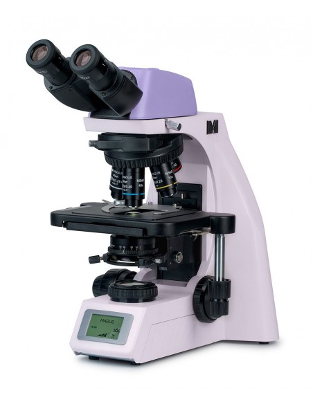
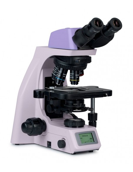
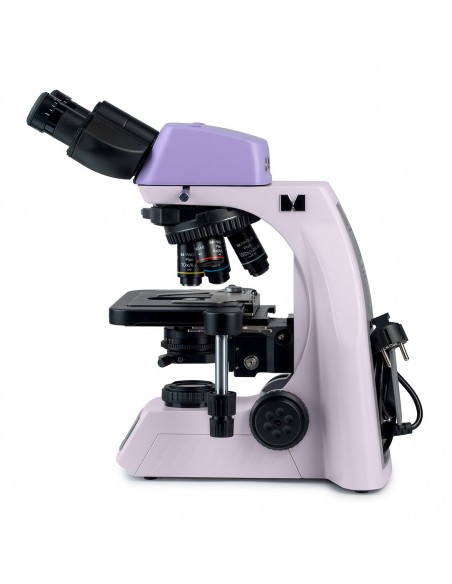
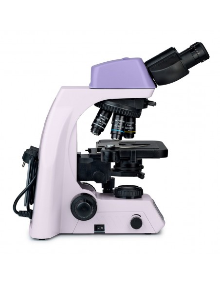
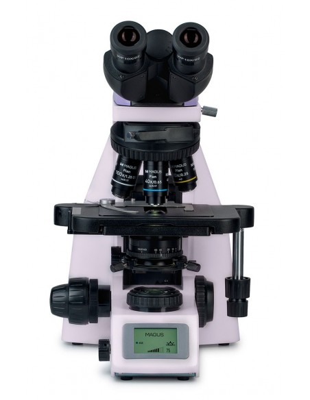
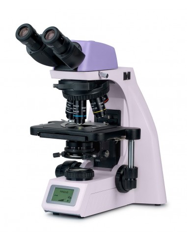
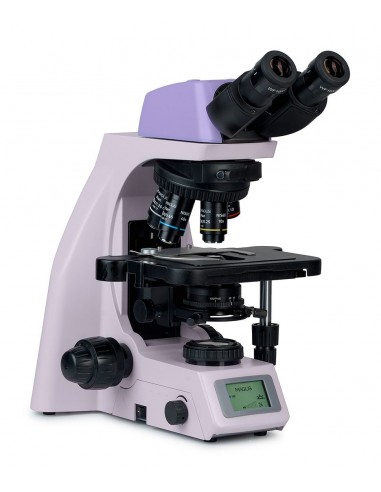
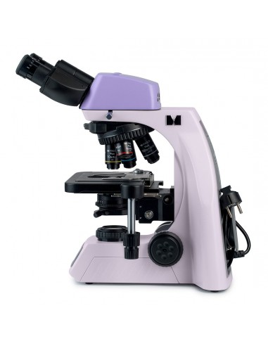
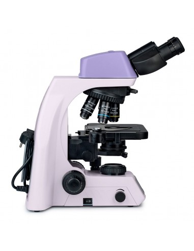
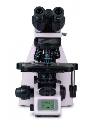
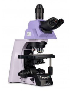
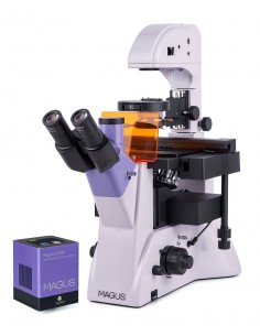
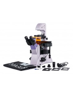
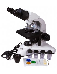
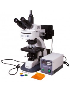
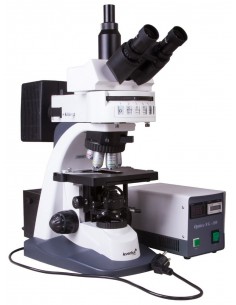
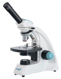
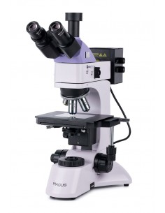
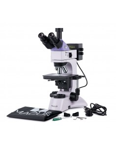
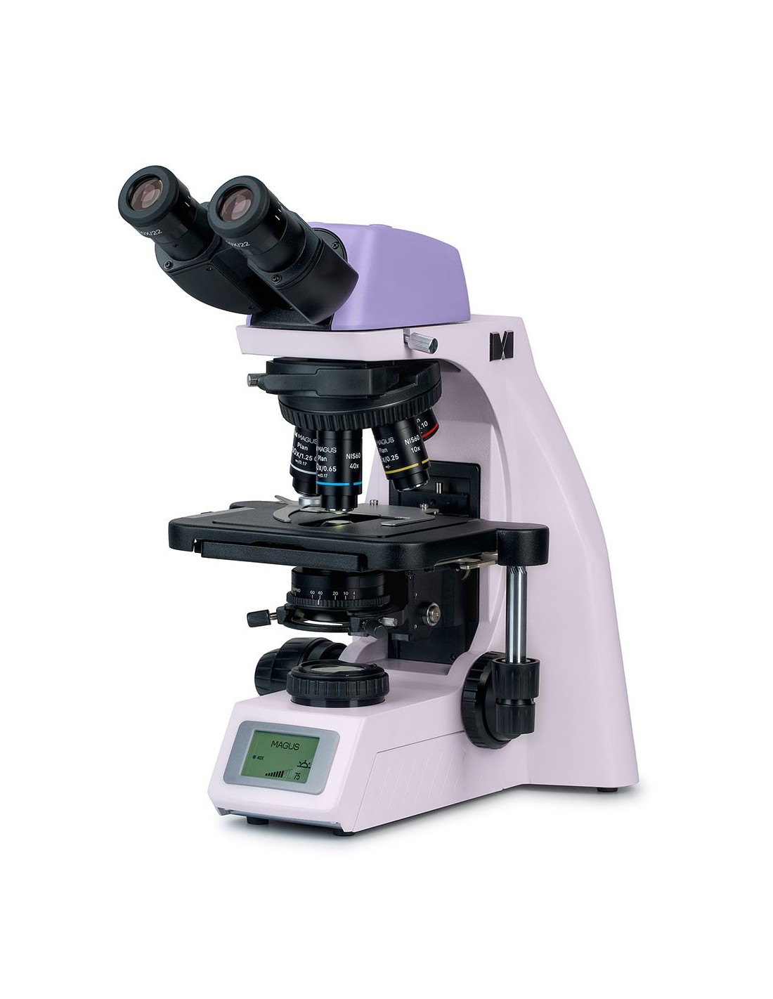
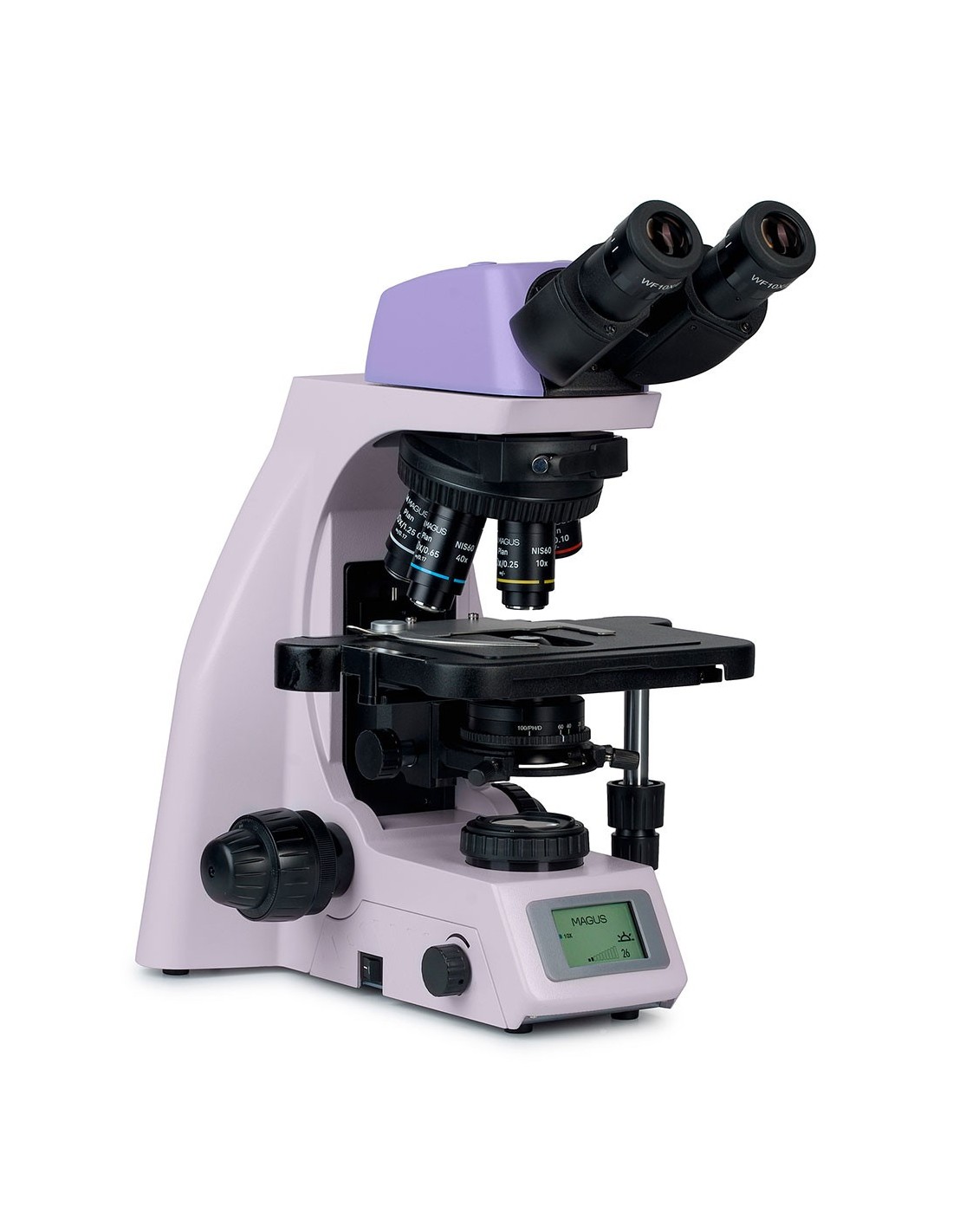
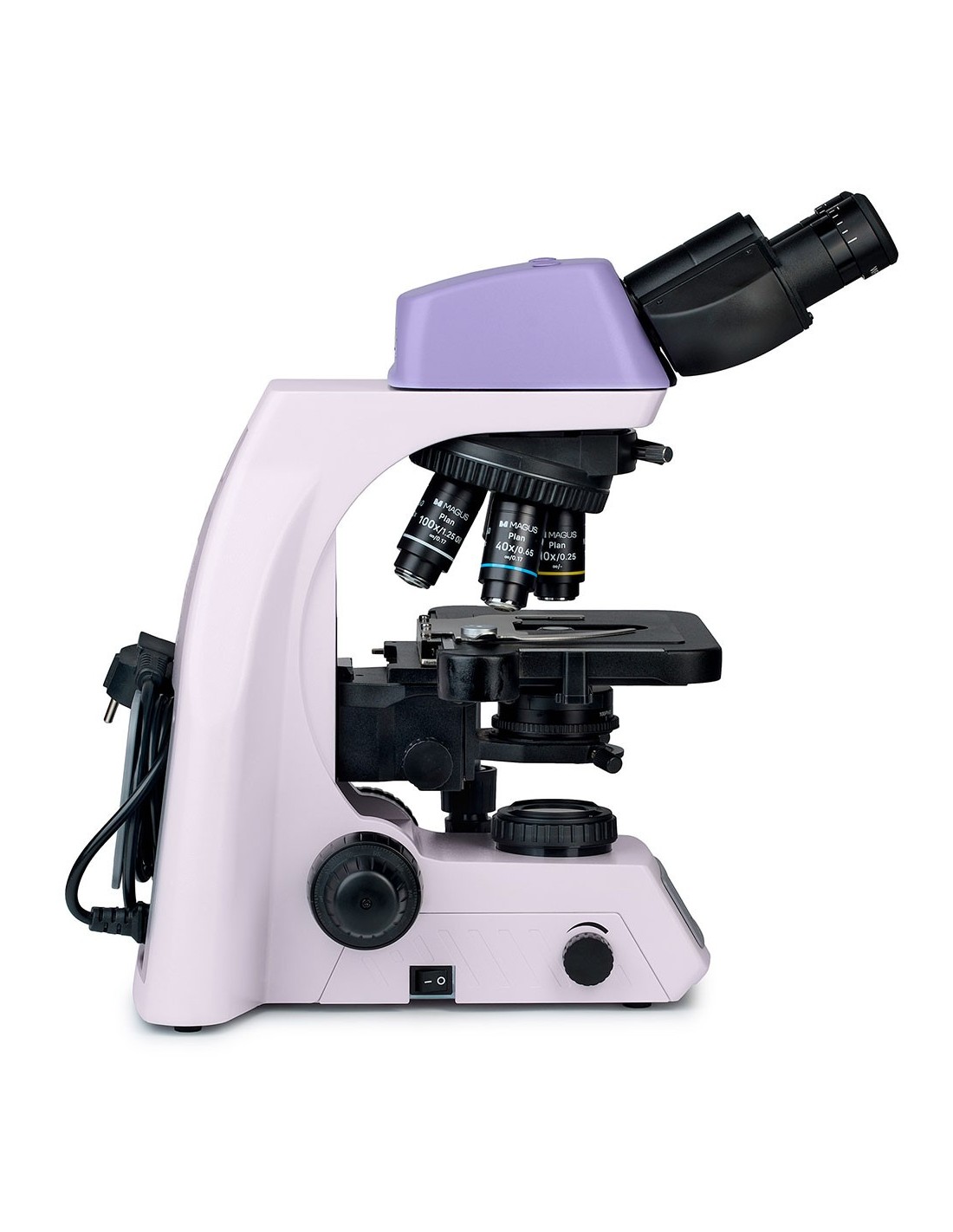
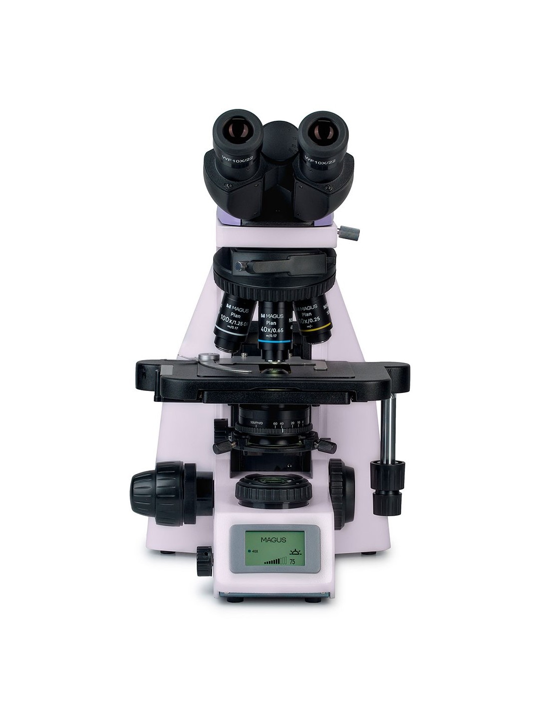
The MAGUS Bio DH260 microscope is a microscope for everyday use designed in the basic configuration to work with biological samples in transmitted light using the bright field method. Equipping the device with additional components will allow you to conduct research in fluorescent or polarized light, as well as use the dark field or phase contrast method. The microscope features a binocular head with integrated camera and a coded nosepiece that, when changing magnification, maintains the illumination intensity selected by the user. It also features an LCD screen that displays the operating parameters of the microscope.
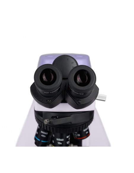
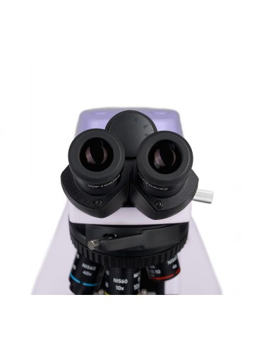
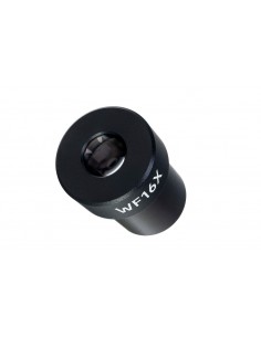
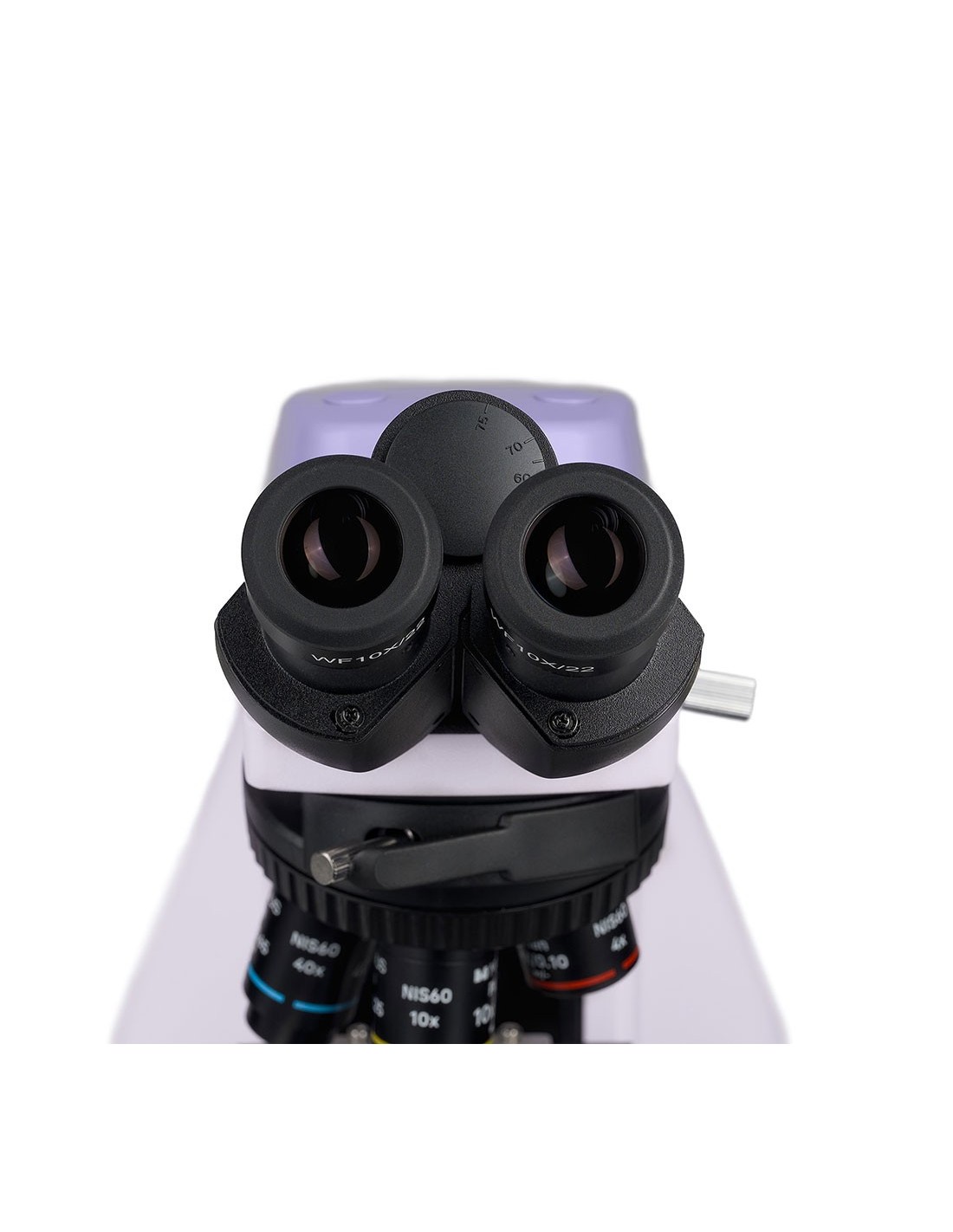
The head with two eyepiece tubes is complemented by a digital camera. The eyepiece tubes are designed for infinity optics. The microscope kit includes 10x/22mm eyepieces. which are equipped with a mechanism for diopter adjustment and a long eye relief for working while wearing glasses. To protect the lenses from rubbing and scratches, rubber eyecups are placed on the eyepieces.
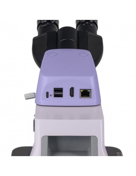
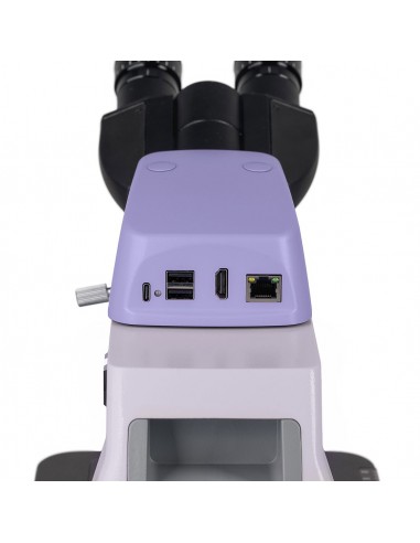
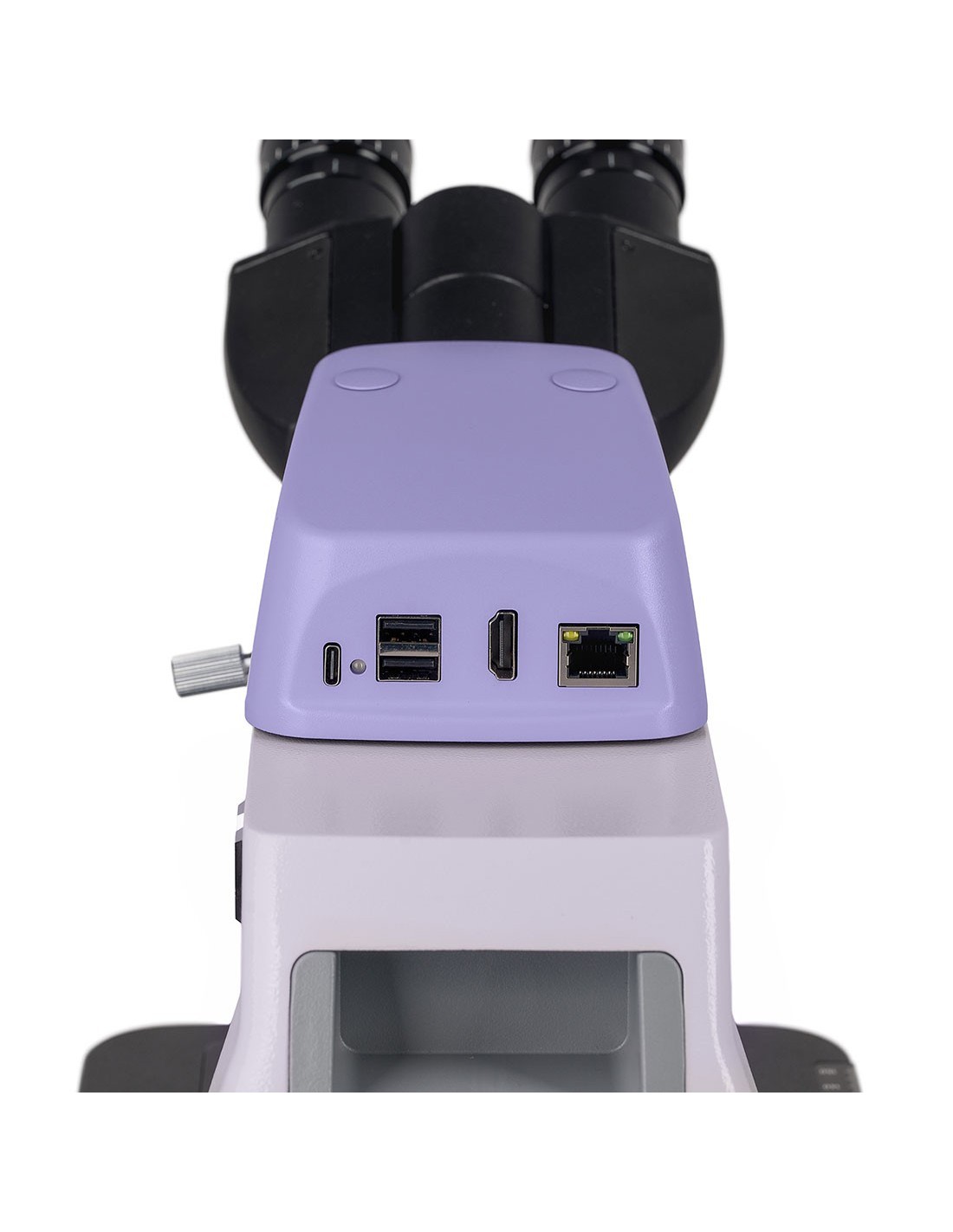
The integrated digital camera with 8 MP sensor produces images with 4K resolution (3840x2160 px). The high resolution will allow you to study the sample in detail. The camera is most effective when paired with 4x and 10x lenses. When shooting video, the camera produces 30 fps at maximum resolution - the footage is high quality and movement is conveyed authentically with no "gaps" between frames. The camera can be used for recording photos and video searches, for displaying images on the screen and for focusing the optical system. The device is equipped with a Wi-Fi interface that is used to transfer data to external devices.
The nosepiece has slots for 5 lenses. It is installed "inwards": the working target is located closest to the user, and the other targets are directed in the opposite direction. This position leaves a lot of free space in front of the user, which can be used to manipulate the sample. The microscope is equipped with four planachromatic objectives of different magnifications; user can fill the fifth slot according to their needs. The parfocal distance is 60 mm. The housing above the nosepiece is designed to accommodate the analyzer when working in polarized light.
The nosepiece is coded: the user sets the light intensity for each objective once and then, when you turn the revolver, the light is automatically adjusted. This feature makes working under the microscope faster and more comfortable: each objective transmits light differently depending on its magnification. When changing objectives, the intensity of the light in the eyepiece changes significantly and each time it is necessary to adjust it. Additionally, changes in light brightness tire the eyes. The coded revolver saves time and effort.
Focusing is carried out using the coarse and fine adjustment knobs which are located on the same axis. The coarse adjustment is carried out with the handle on the left, while for the micrometric adjustment there are handles on both sides. The stiffness of the coarse focus travel can be adjusted with the special adjustment ring on the left.
The stage does not have a positioning rack, which makes the design more ergonomic. The sample moves along the stage surface using a belt drive mechanism. The sample holder is initially attached to the stage with two screws, but can be quickly removed if manual scanning is performed. The stage is controlled by a long handle: to manipulate it, the user can comfortably keep his hands on the table.
The condenser under the stage controls the illumination of the sample using an iris diaphragm. For ease of adjustment, the condenser on this microscope is equipped with markings indicating the magnification of the objectives, and on its ring there is a pointer, set to the mark corresponding to the selected objective. As a result, the image is clear and contrasted. When conducting studies using dark field or phase contrast methods, the corresponding sliders are installed in a housing on the condenser.
The light source in the microscope is a 3 W LED with a color temperature stable. The LEDs have a lifespan of 50,000 hours. The microscope is equipped with Köhler illumination, which creates uniform illumination of the object across the entire field of view. The objectives operate at maximum resolution and dust or other foreign objects do not enter the field of view.
The operating parameters are displayed on the LCD screen which is integrated into the base of the microscope. You can view the selected objective magnification, illuminator brightness and operation mode, and set the auto shut-off time. Manipulations are performed using two handles.
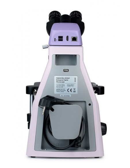
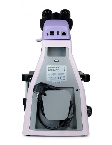
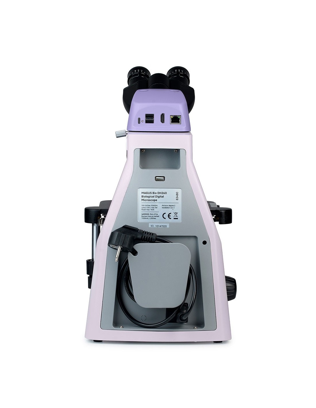
The design of the microscope has been well thought out to ensure comfortable work. There is a special handle to move it, and the power cord is hidden - it is safe and convenient for operation and storage. The microscope can be integrated with the necessary accessories to expand its functionality: eyepieces, components for optional research methods and a calibration slide for measuring objects.
Key features:
- The microscope works with biological samples in bright-field transmitted light. If equipped with additional accessories, dark field, phase contrast, luminescence or polarized light methods become available
- The head with two tubes is completed with an integrated digital camera; the position of the tubes is height-adjustable
- The five-objective revolver "remembers" the illumination settings for each magnification
- The condenser is centered and height-adjustable; has magnification marks to adjust the aperture position for each objective
- You can adjust the illumination using the Köhler method
- The LCD screen at the bottom of the microscope displays the operating parameters
- Ergonomic design: features a carrying handle and hidden power cord
The package contains:
- Support with integrated power supply, transmitted light source, focusing mechanism, stage, condenser mount and nosepiece revolver
- Abbe NA 1.25 condenser oil
- Binocular head with camera integrated digital
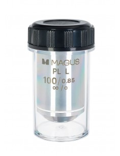
- Plan achromatic objective with infinity correction: 4x/0.10, parfocal height 60 mm
- Plan achromatic objective with infinity correction: 10x/0.25, parfocal height 60 mm
- Plan achromatic objective with infinity correction: 40x/0.65, parfocal height 60 mm
- Plan achromatic objective with infinity correction: 100x/0.25 oil ( with spring mechanism), parfocal height 60 mm
- 10x/22 mm eyepiece with long eye relief and diopter adjustment (2 pcs.)
- Eyecup (2 pcs.)
- Optical filter
- Bottle of immersion oil
- AC power cord
- Dust cover
- User manual and warranty certificate
Available on request:
- 10x/22 mm eyepiece with graduated scale
- 10x/22 mm eyepiece with reticle
- 10x/22 mm eyepiece with viewfinder
- 12.5x/17.5 mm eyepiece (2 pcs.)
- Eyepiece 15x/16 mm (2 pcs.)
- Eyepiece 20x/12 mm (2 pcs.)
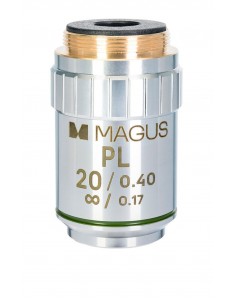
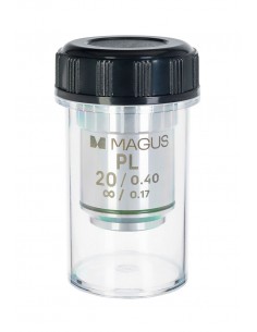
- Planachromatic objective with infinity correction: 20x/0.40, parfocal height 60 mm
- Phase contrast device: phase contrast slider set, auxiliary centering telescope, phase contrast objective set
- Dark field slider li>
- Polarizer tool
- Calibration glass
| Brand | MAGUS |
| Warranty | 5 лет |
| EAN | 5905555019468 |
| Package size (LxWxH) | 47x32x67 cm |
| Shipping weight | 12.1 kg |
| === Delimiter === | Microscope Specifications |
| Type | biological, light/optical, digital |
| Head | binocular |
| Vertical tube | Gemel head (Siedentopf type, with 360° rotation) |
| Head inclination angle | 30° |
| Magnification, x | 40–1000 in basic configuration (*optional: 40–1250/1500/2000) |
| Range magnification | from 800x to 1280x |
| Eyepiece tube diameter, mm | 30 |
| Eyepieces | 10x/22, long eye relief (*optional: 10x/22 with scale, 10x/22 with reticle, 10x/22 with viewfinder, 12.5x/17.5, 15x/16, 20x/12) |
| Objectives | planachromatic, infinity corrected: 4x/0.10; 10x/0.25; 40x/0.65; 100xs/1.25 oil (*optional: 20x/0.40); parfocal height 60 mm |
| Nosepiece revolver | 5 objectives, coded |
| Working distance, mm | 30 (4x); 10.2 (10x); 1.5 (40x); 0.2 (100xs) |
| Interpupillary distance, mm | 47 — 78 |
| Stage, mm | 230x150 |
| Range of movement of the stage, mm | 78/54 |
| Stage features | two-axis mechanical translator, without positioning rack |
| Eyepiece diopter adjustment, diopters | ±5D on each eyepiece |
| Eyepiece diopter adjustment | yes |
| Condenser |
| Condenser |
| td> | centrable and height-adjustable Abbe condenser NA 1.25 with adjustable aperture diaphragm and housing with socket for dark field slider and phase contrast, dovetail mount |
| Focus | coaxial, macrometric ( 30 mm, 37.7 mm/rev, with tension adjustment mechanism) and micrometric (0.002 mm, 0.2 mm/rev) |
| Illumination |
| Brightness adjustment | yes |
| Power | 100–240 V, 50/60 Hz, AC mains |
| Type of light source | 3 W LED |
| Operating temperature range, °C | 0 — 70 |
| Extra elements | automatic brightness adjustment when changing lenses, status display on LCD screen, standby mode, eco mode |
| Possibility to connect additional equipment | dark field sliders, polarization instruments (polarizer and analyzer), phase contrast devices (sliders and objectives) |
| User level | expert users, professionals |
| Assembly and installation level of difficulty | complicated |
| Application | laboratory/medical |
| Lighting position | lower |
| Search method | bright field |
| Digital camera included | yes |
| Set with pencil case/case/bag | dust cover |
| === Delimiter === | Digital camera |
| Color/monochrome | color |
| Megapixel | 8 |
| Maximum resolution, px | 3840x2160 |
| Sensor size |
| Sensor size |
| td> | 1/1.8" |
| Pixel size, µm | 2x2 |
| Interface connectors | Wi-Fi |
| Video recording | yes |
| 30 |
| Installation location | integrated |
| Camera power | main cable |