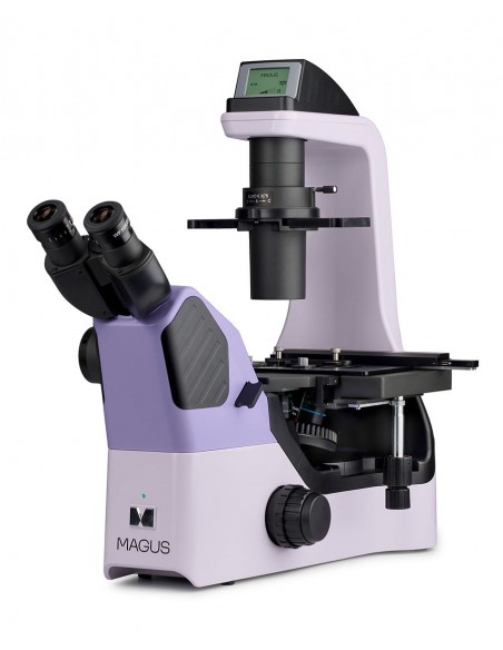
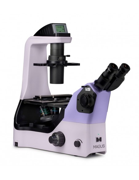
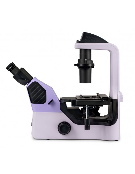
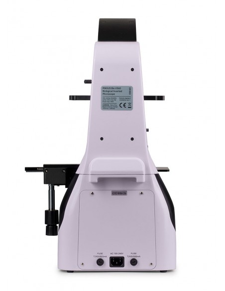
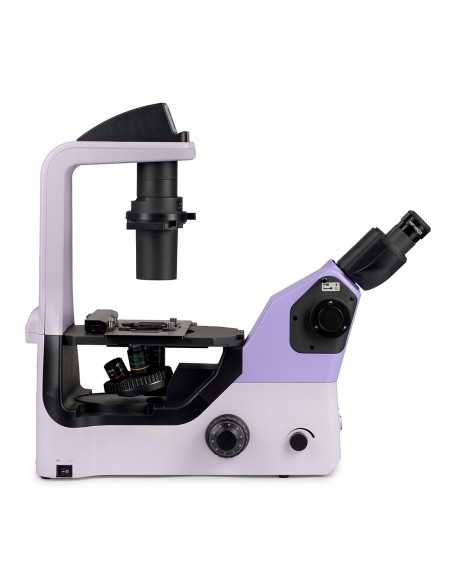
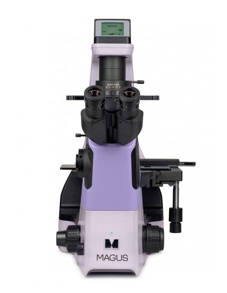
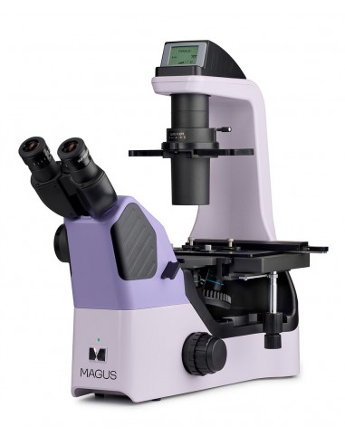
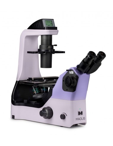
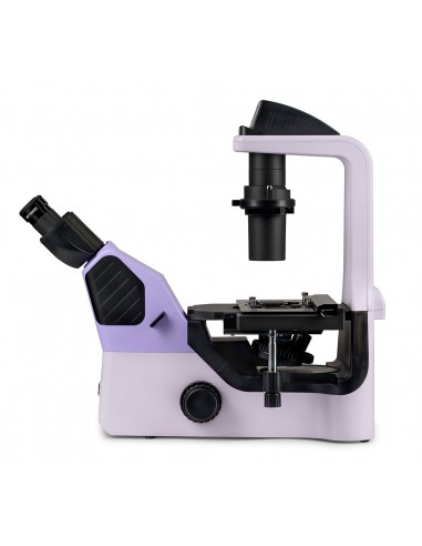
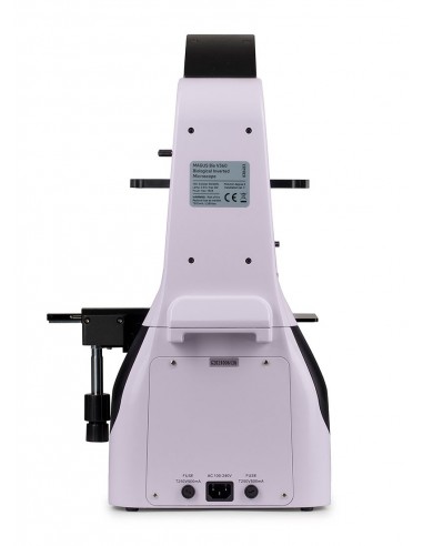
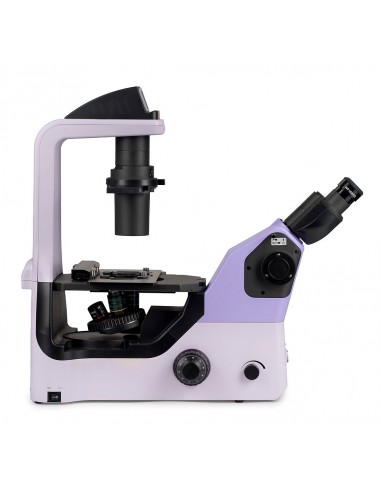
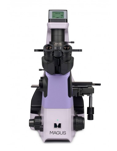
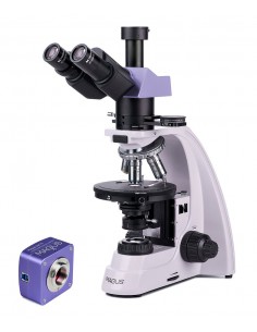
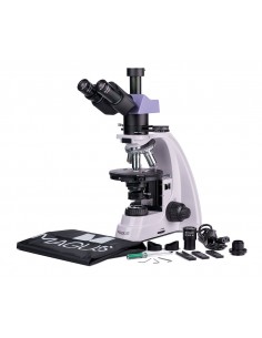
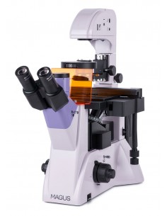
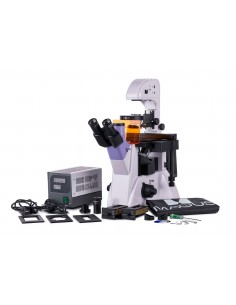
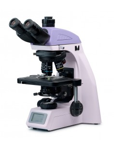
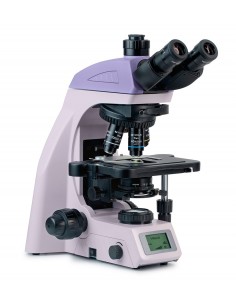
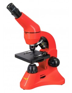
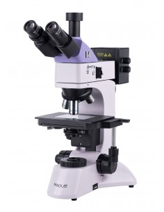
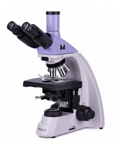
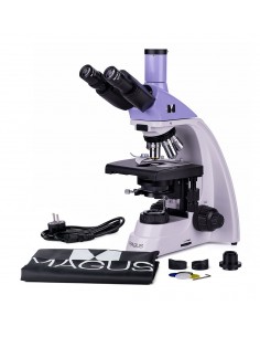
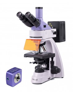
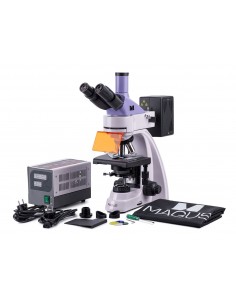
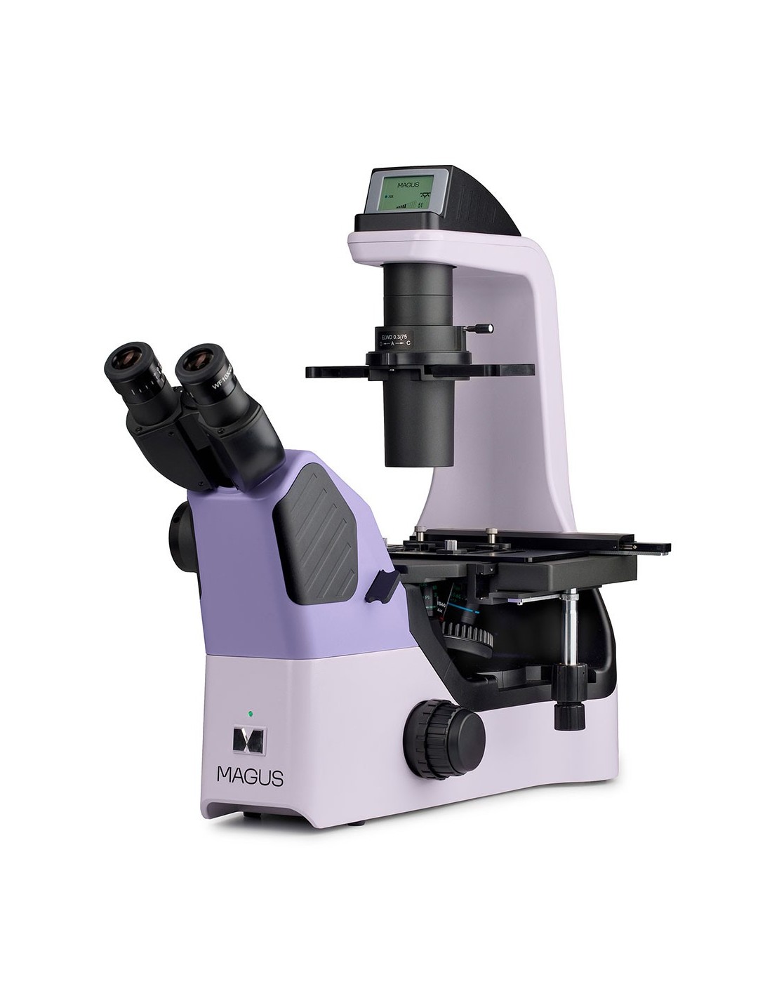
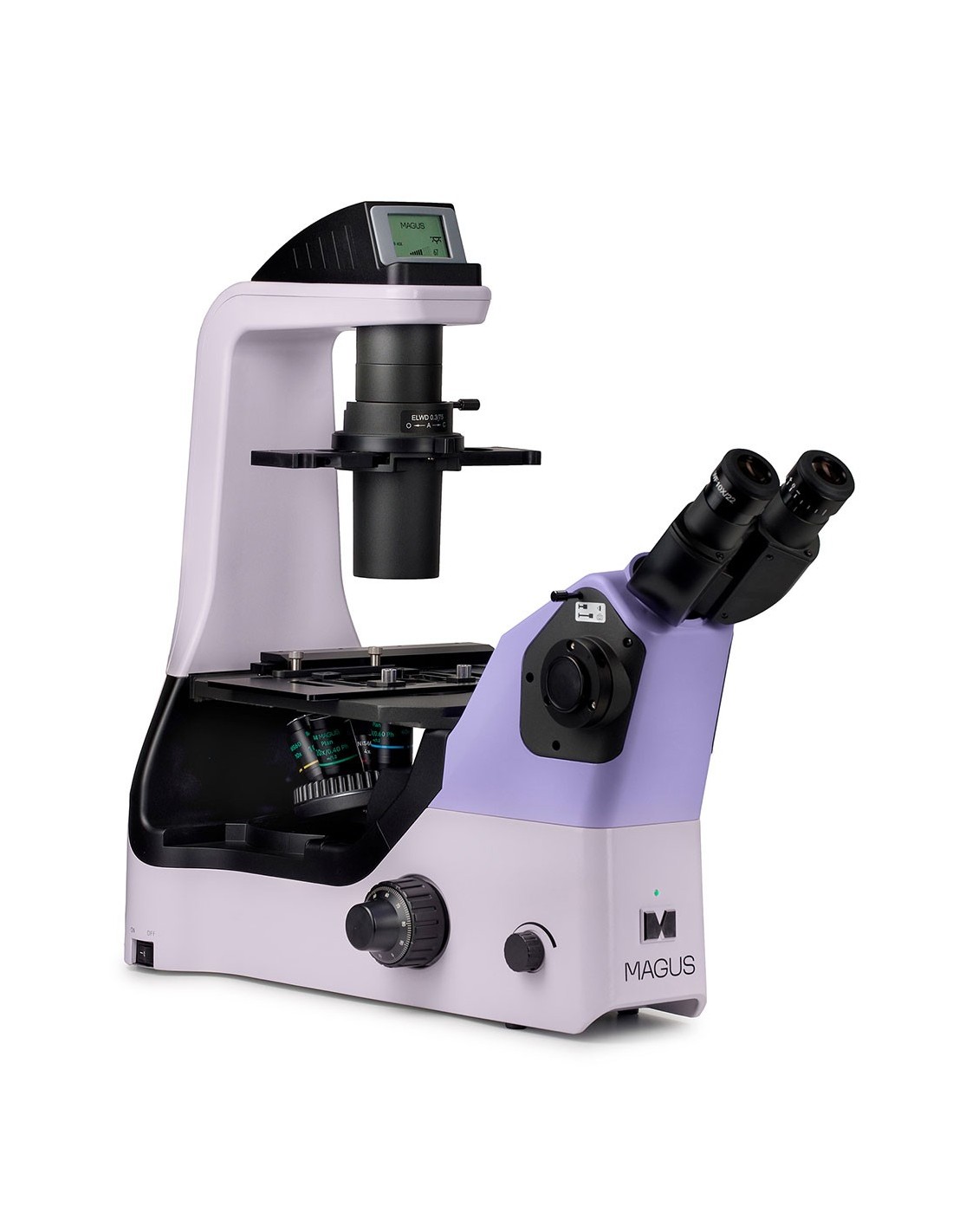
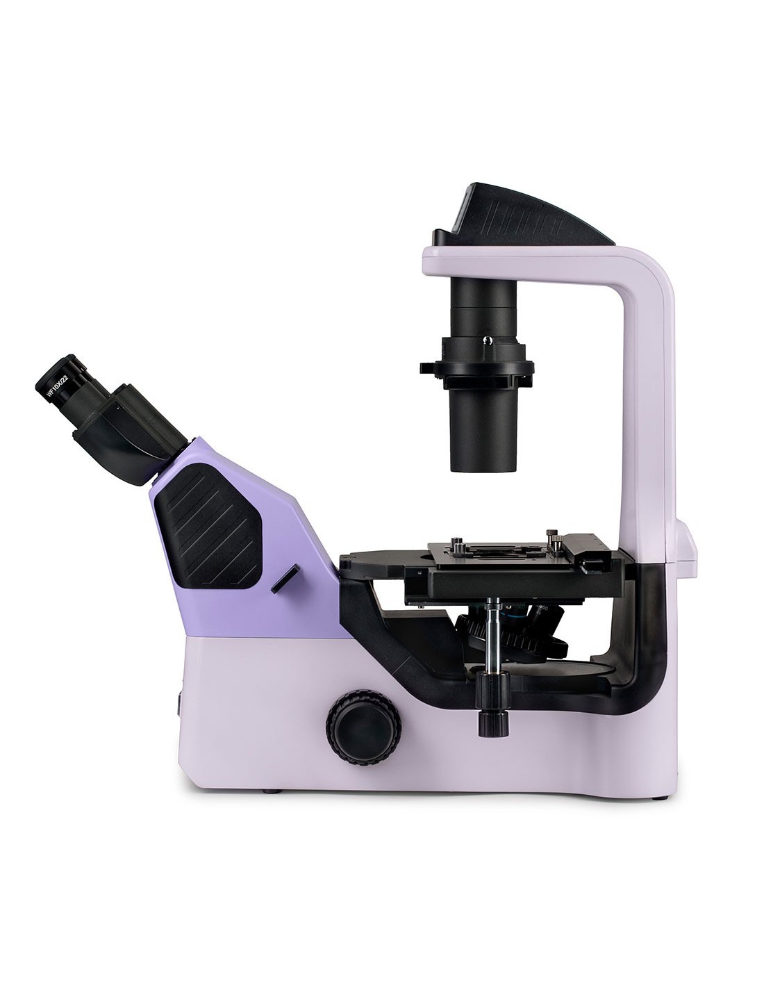
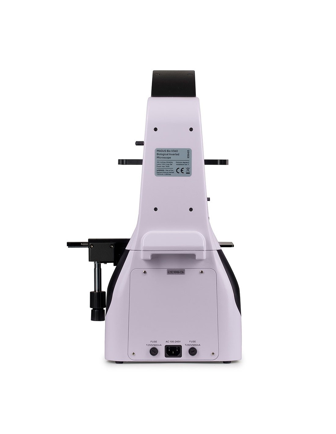
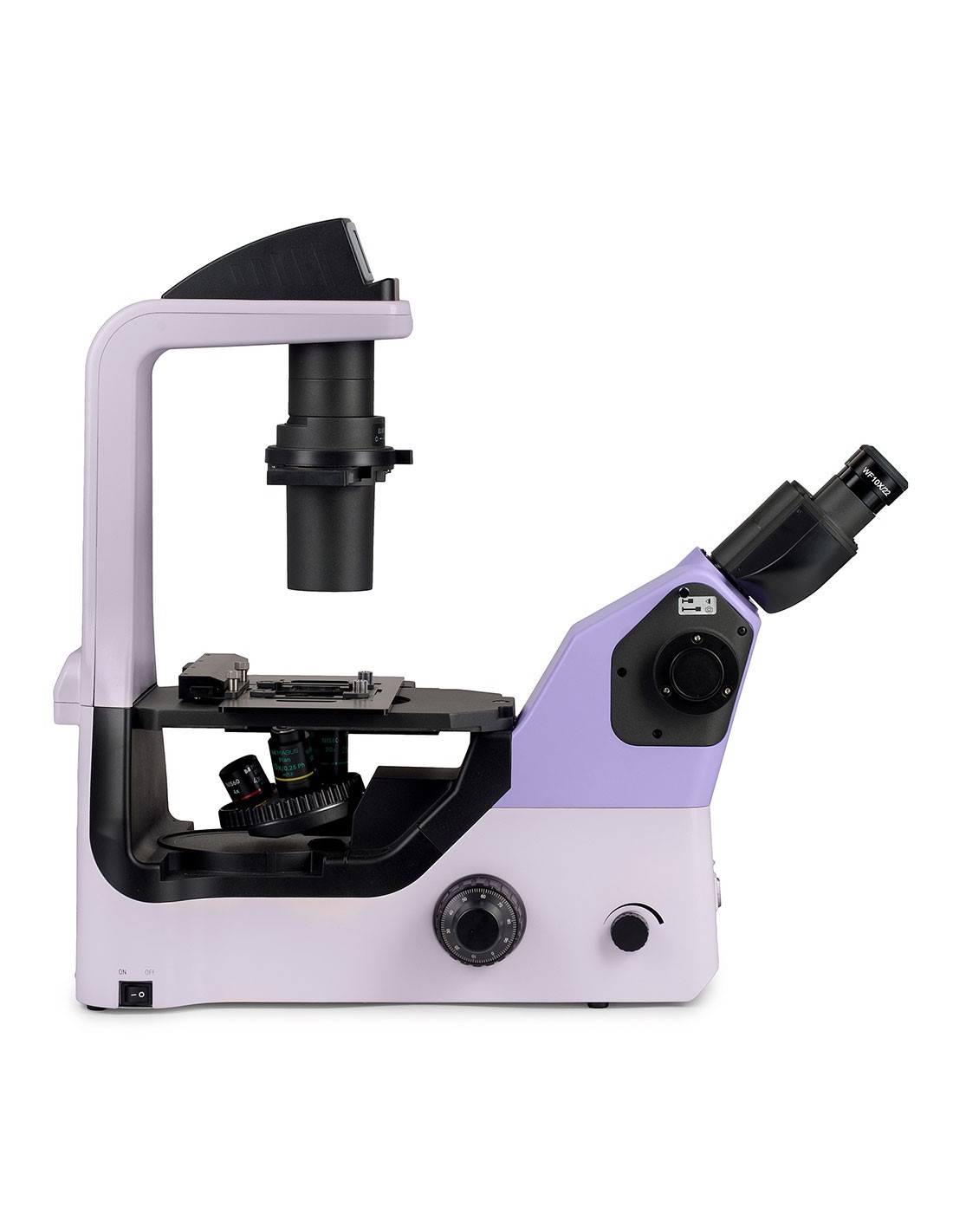
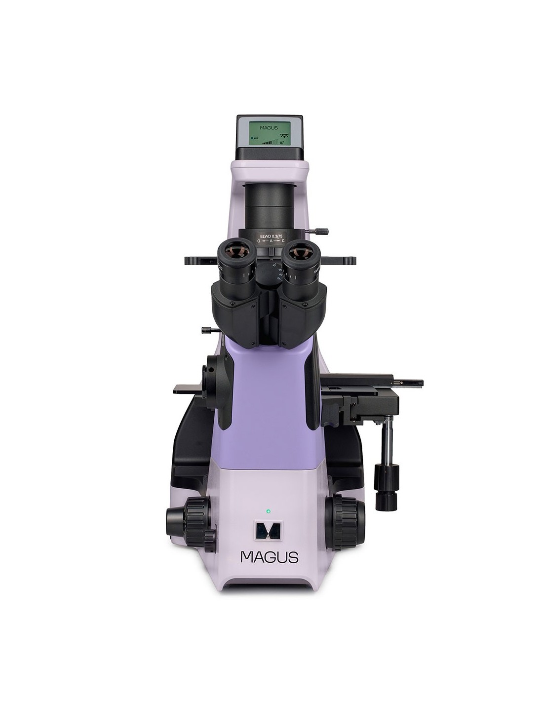
Research microscope.
Designed for the study of liquid precipitates, cell colonies, live cells, tissue cultures and other stained and unstained specimens in laboratory containers.
Inverted design allows the use of plates Petri dishes, multi-well plates, vials, roller bottles and flasks up to 75 mm and with a 1.2 mm thick bottom. The microscope uses special objectives for observation with such containers.
By removing the condenser, cell colonies can be observed in Petri dishes or in cylindrical flasks up to 187 mm.
The technique of transmitted light microscopy ( brightfield and phase contrast) is used for the study of samples.
The installation of optional components allows you to use the Hoffman modulation contrast and relief contrast methods.
The microscope is used for research in medicine, pharmacology, biology and virology. The highest configuration of the microscope is suitable for IVF.
Microscope head
Binocular head with infinity corrected optics. The digital camera is installed in the side port on the microscope head. Beam division of 100/0 or 0/100.
Nosepiece nosepiece
Nosepiece nosepiece coded for 5 objectives. Alternatively, an additional objective can be installed in the free slot to obtain extra magnification.
Objectives
Infinity corrected plan achromatic objectives with long focusing distance work for plates with a thickness of less than 1.2 mm. The parfocal distance is 60 mm.
To work using the bright field method, one objective is used, while to work using the phase contrast method, three objectives are used.
Maintaining comfortable brightness levels when magnification is changed
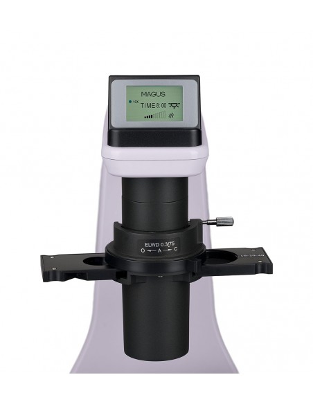
Objectives with different magnifications transmit light at different levels of intensity; every time you change lenses, you need to adjust the brightness of the light. Additionally, brightness increases dramatically when switching from a higher magnification objective to a lower magnification objective. The strong increase in brightness makes the eyes tiring. MAGUS Bio V360 is equipped with intelligent brightness control. The microscope remembers the brightness of each objective that the user has selected and automatically sets it when the nosepiece is moved. Intelligent control reduces the time needed to adjust brightness. MAGUS Bio V360 increases user comfort and saves time even when the task requires frequent changes of magnification.
Focusing mechanism
The coarse and fine focus knobs are coaxial and positioned at the bottom. The researcher can rest his hands on the table and take a comfortable position in front of the microscope.
The coarse focus lock knob helps to quickly adjust the microscope after changing the study object. The knob is located on the left side of the microscope on the same axis as the focusing mechanism.
The ring on the right side adjusts the tension of the coarse focus screw. The user adjusts the right tension for the observations.
Stage
The mechanical stage is fixed. A special mechanism is installed on the table that moves the laboratory containers along two directions perpendicular to each other. The fluid and micrometric movement of the object offers an accurate study: no part of the sample will be overlooked.
The long control handle of the stage guarantees comfortable observations: the hand can rest on the table without effort.
The Microscope kit includes a universal plate holder.
Condenser
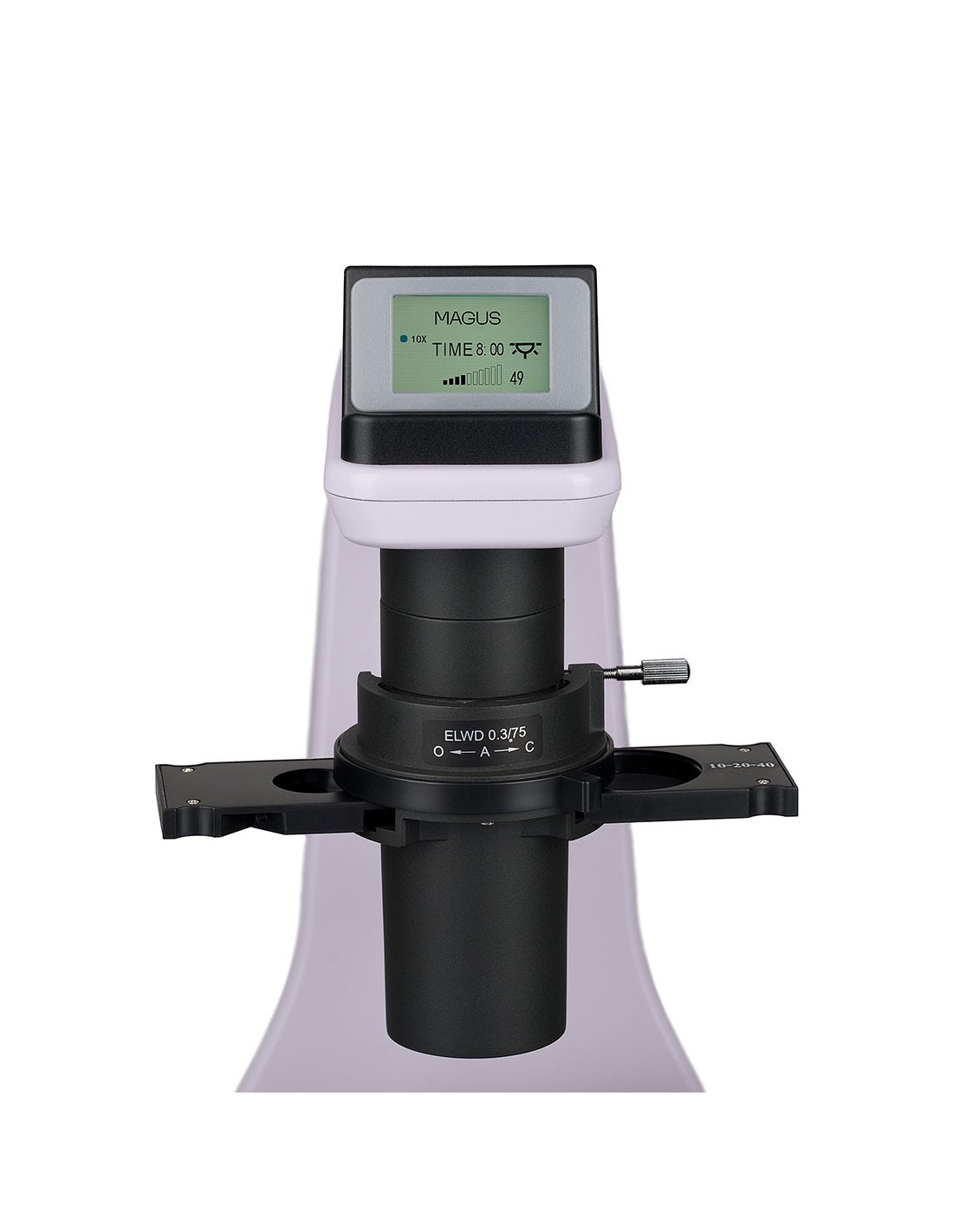
A phase contrast slider is installed in the condenser. You can center the phase contrast rings. Using the slider saves time when switching from one microscopy technique to another.
A contrast relief or Hoffman modulation contrast slider can be installed in the condenser slot.
Illumination
The 3W LED provides bright illumination, sufficient for brightfield and phase contrast techniques with all types of objectives. The color temperature does not change when the brightness is adjusted. The life cycle of the LEDs is 50,000 hours.
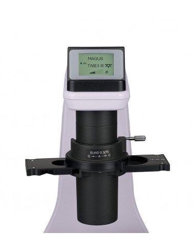
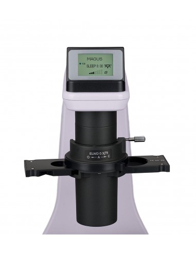
LCD status screen
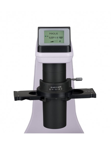
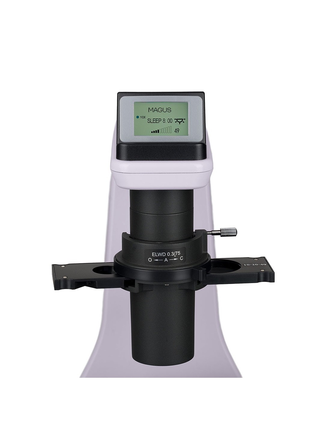
The LCD screen at the base of the microscope shows the magnification of the objective, the brightness of the light source and the operating mode (“sleep”, “eco”).
Using the screen and two knobs, the user adjusts the brightness, locks the brightness adjustment and sets the standby and the automatic sleep timer.
Ergonomic design
Physical discomfort causes fatigue and reduces productivity. The ergonomic design of the microscope plays an important role in daily scientific research.
MAGUS Bio V360 allows the user to carry out his task comfortably. The microscope head is positioned at an ergonomic angle so that the back and neck do not get tired.
Thanks to the low, compact stage, the user can handle the sample and work with laboratory containers in a practical way .
The long handle of the movement mechanism ensures that it is neither necessary to lift the hand from the table to control the microscope nor to change the position of the body.
The focus knobs are positioned at the base of the microscope. The user does not need to strain his hands. Thanks to the smooth movement of the mechanism, the user can focus on the object without problems.
Accessories
There is a line of accessories designed for this microscope. Eyepieces extend the magnification range of the microscope. Additional eyepieces help to exploit the full potential of the most often used objective.
Hoffman modulation contrast and relief contrast devices extend contrast techniques and allow the analysis of non-visible objects in bright field and phase contrast.
A digital camera plays the microscope image onto a monitor and stores the files, while software acquires sample measurements in real time.
A calibration slide can be used to measure objects and combine it with an eyepiece with a scale or camera software.
Key features:
- Analysis of colored objects and not in laboratory containers: Petri dishes, flasks, plates, work in containers with a height of up to 185 mm
- Research methods: bright field and phase contrast; with additional equipment: Hoffman modulation contrast and relief contrast methods
- Coded nosepiece: the brightness of the light source is automatically set according to the selected objective
- Binocular head with side tube for digital camera; beam split of 100:0 or 0:100
- The transmitted light illuminator is an energy-saving 3W LED with a lifespan of up to 50,000 hours
- Capacitor with slot for installation of sliders; phase contrast slider included, centered phase rings
- Intelligent lighting control system: automatic brightness selection, light dimming lock, automatic shutdown with timer, LCD status screen
- Stage with movement of containers along the package contains:
- Support with integrated power supply, transmitted light source, focusing mechanism, stage, condenser mount and nosepiece
- Condenser with slot for slider
- Binocular microscope head with side tube
- Infinity corrected plan achromatic objective: 4x/0.10 WD 30 mm, parfocal height 60 mm
- Infinity corrected plan achromatic objective: 10x/0.25 phase WD 10.2 mm, parfocal height 60 mm
- Infinity corrected plan achromatic objective: 20x/0.40 phase WD 12 mm, parfocal height 60 mm
- Infinity corrected plan achromatic objective: 40x/0.60 phase WD 2.2 mm, parfocal height 60 mm
- 10x/22 mm eyepiece with long eye relief and adjustment of the diopter (2 pcs.)
- Eyecup (2 pcs.)
- Centering telescope
- Phase contrast slider with phase contrast rings centering
- Round stage plate
- Universal plate holder
- C-mount adapter
- AC mains power cable
- Dust cover
- User manual and warranty card
Available upon request:
- Eyepiece 15x/16 mm (2 pcs.)
- Eyepiece 20x/12 mm (2 pcs.)
- Device for analysis using the Hoffman modulation contrast method < li>Device for analysis using a relief contrast method
- Digital camera
- Calibration slide
- Monitor
| Brand | MAGUS |
| Warranty | 5 лет |
| Package size (WxDxH) | 56x65x67 cm |
| Shipping weight | 24.4 kg |
| Type | biological, light/optical |
| Head | binocular |
| Vertical tube | beam split 0/100, 100/0 , Siedentopf, with side tube |
| Head inclination angle | 45° |
| Magnification, x | 40–400 in the basic configuration (*optional: 40–600/800) |
| Magnification range | from 200x at 800x |
| Eyepiece tube diameter, mm | 30 |
| Eyepieces | 10x/22 mm, long eye relief (*optional: 15х/16 mm, 20х/12 mm) |
| Lenses | infinity plan achromatic objectives : 4x/0.10; 10x/0.25 phase; 20x/0.40 phase; 40x/0.60 phase; parfocal distance: 60 mm |
| Nosepiece revolver | 5 objectives, coded |
| Interpupillary distance, mm | 48 — 75 |
| Coffee table, mm | 250x170 |
| Features fixed stage | , with mechanical device for sample movement and 110 mm diameter glass insert, universal support for plates |
| Dioptric adjustment of the eyepiece, diopters | ±5D on each eyepiece |
| Diopter adjustment of the eyepiece | yes |
| Condenser | NA 0.3, working distance: 75 mm; with adjustable aperture diaphragm and a slot for contrast relief, phase or modulation slider |
| Diaphragm | adjustable aperture |
| Lighting | LED |
| Brightness adjustment | yes |
| Power supply | AC mains, 85–265 V, 50/60 Hz |
| Type of light source | LED 3 W |
| Operating temperature range, °C | 10 — 35 |
| Extra elements | automatic brightness adjustment when changing lenses, status display on LCD screen, standby mode, eco mode |
| Special features | 10x–40x phase contrast slider, auxiliary centering telescope |
| Possibility to connect additional equipment | device for analysis using the modulation contrast method Hoffman, device for analysis using a significant contrast method |
| User level | expert users, professionals |
| complicated |
| Application | laboratory/medical |
| Research method | bright field, phase contrast microscope |
| Set with pencil case/case/bag | dust cover |