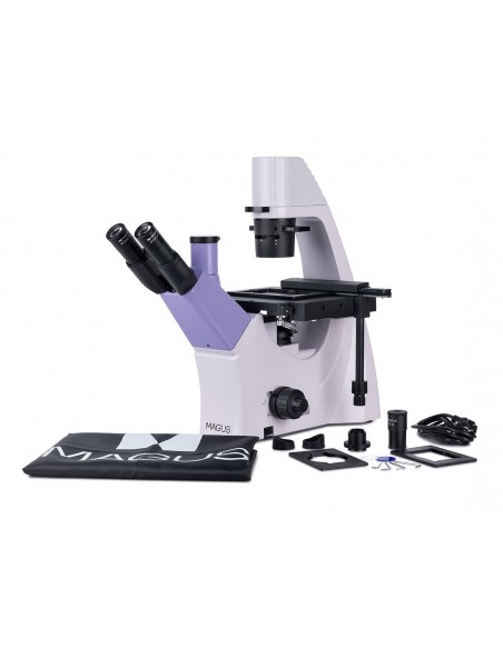
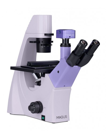
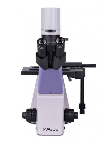
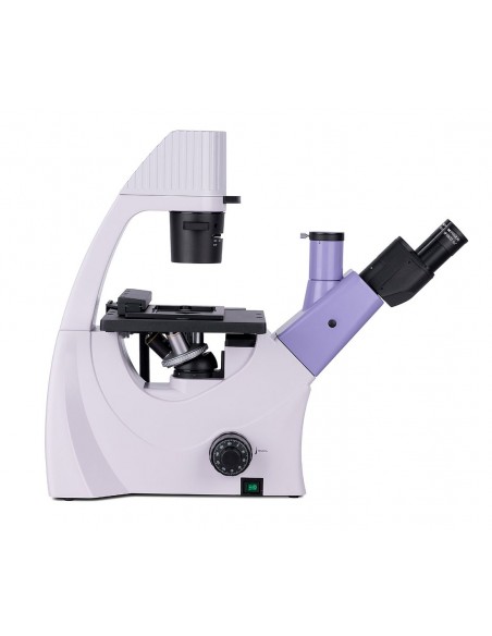
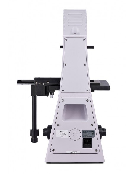
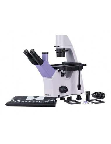
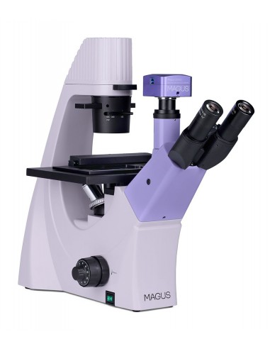
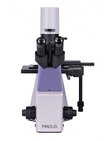
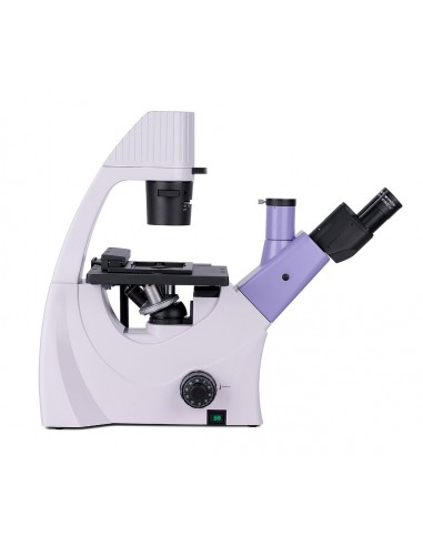
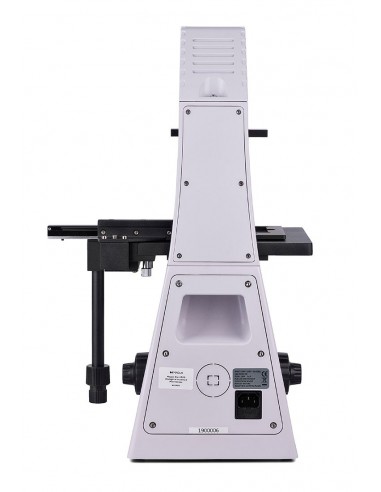
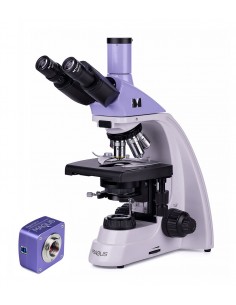
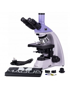
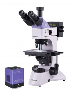
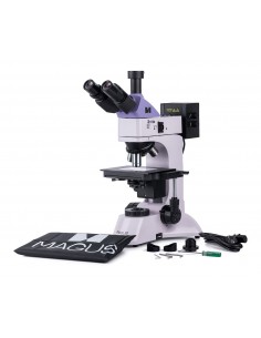
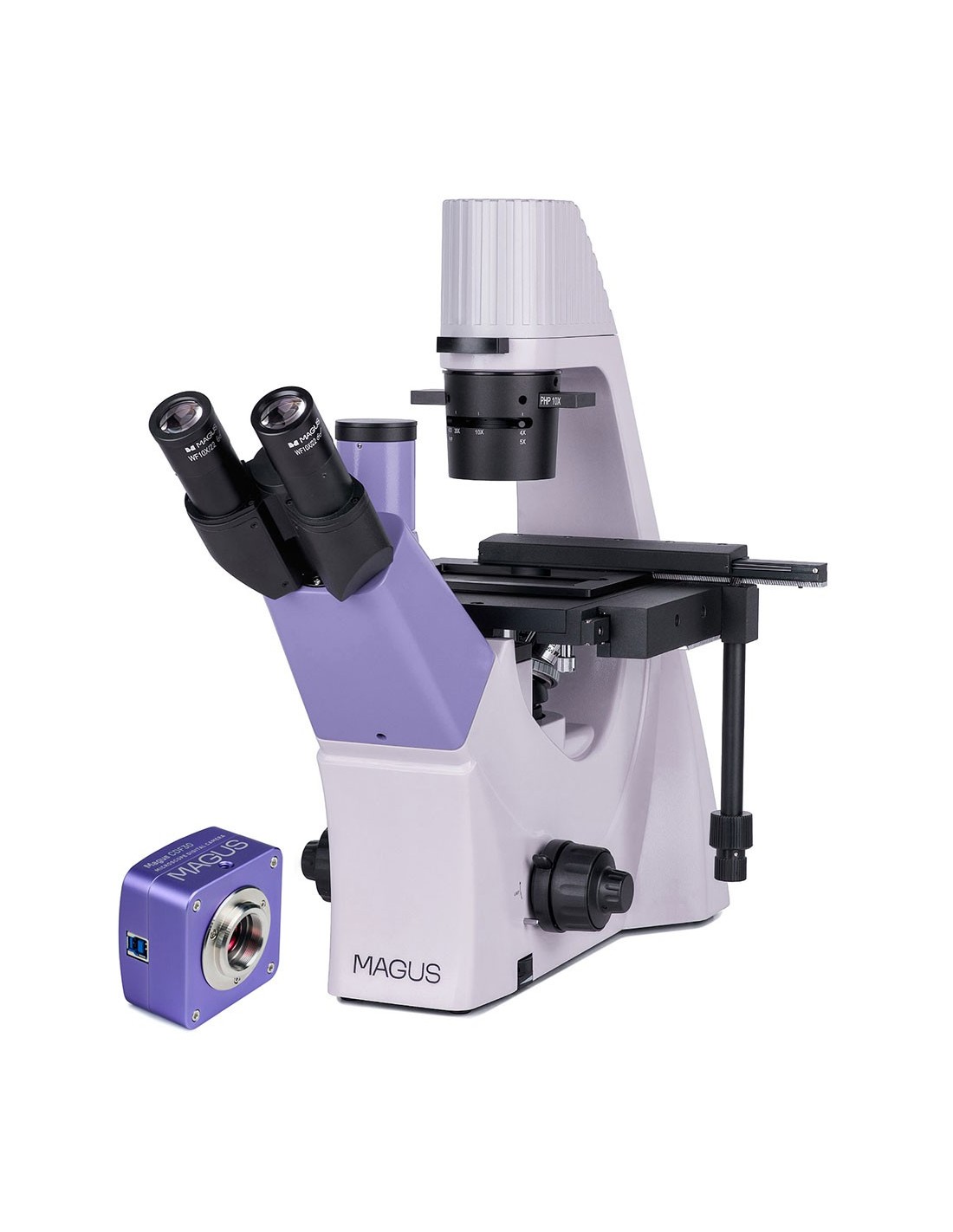
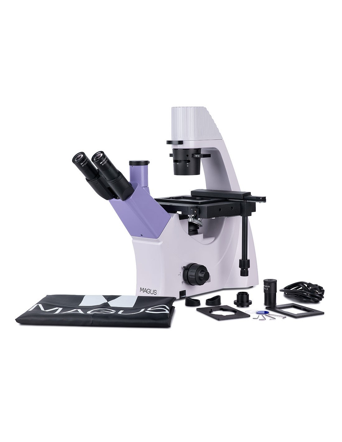
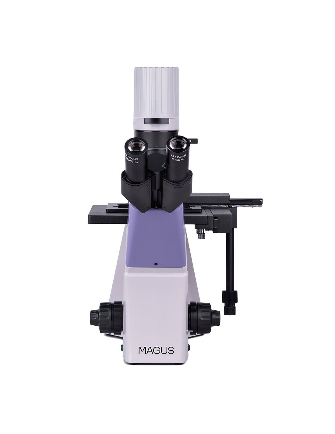
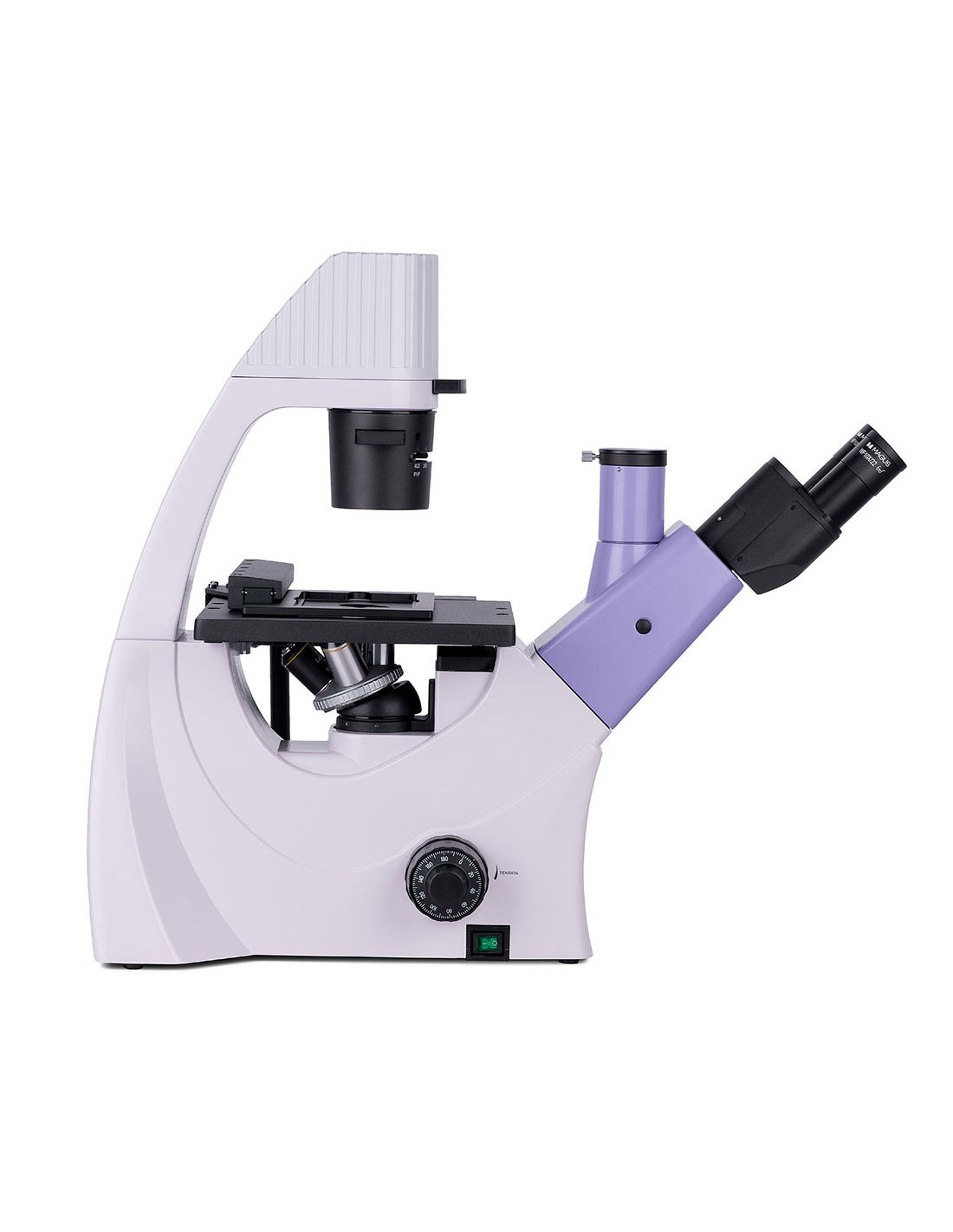
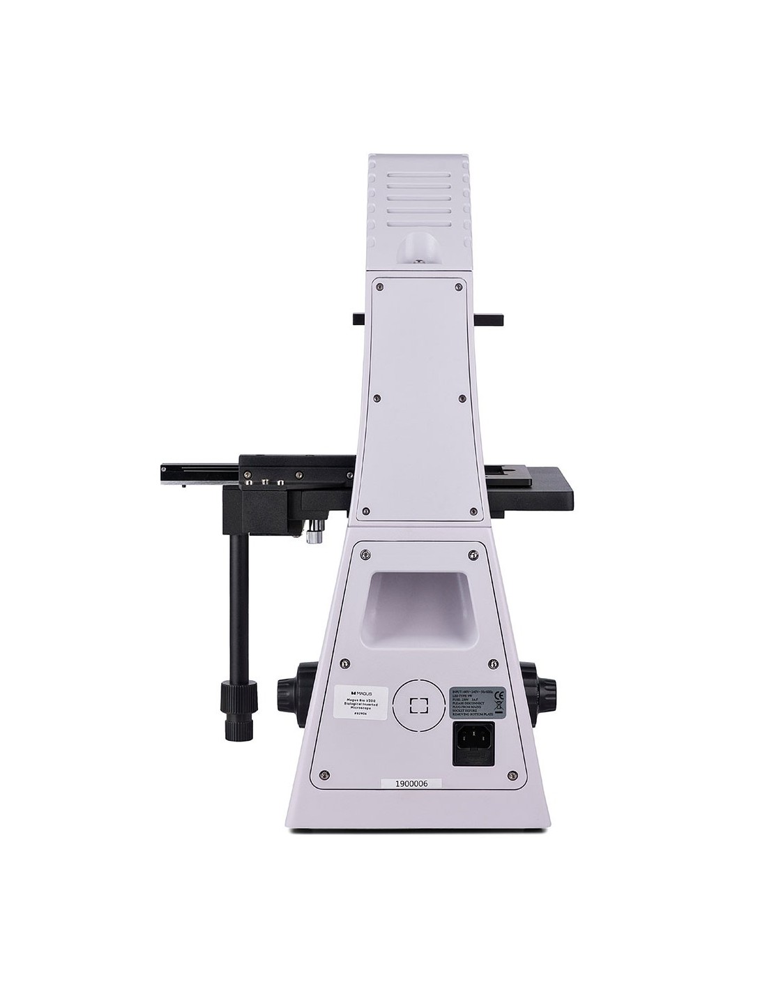
The MAGUS Bio V300 is an inverted design biological microscope for the study of samples on laboratory glassware up to 70 mm in height and with a bottom thickness of up to 1.2 mm. The objects of study can be cell colonies, tissue cultures, biological fluids, etc. Furthermore, the samples do not require the addition of dyes to facilitate examination. The observations are made in transmitted light. Microscopy techniques: bright field and phase contrast. This microscope is suitable for routine work in laboratories and research centers or for teaching.
Digital camera
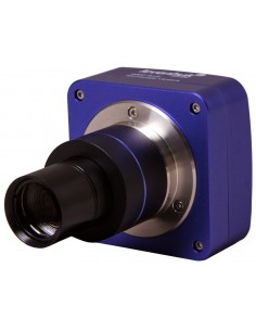
Video optical tube with a sensor digital SONY Exmor 8.3 MP. The sensor uses backlight technology to improve light sensitivity and produce bright, sharp images even in low-light conditions. The camera is used for observation using the bright and dark field microscopy technique on objectives with 4x, 10x and 20x magnification. It can also be used for stereoscopic microscopes. To connect to a computer and display images on the screen, a USB 3.0 port with high data transfer speed (5 Gbps) is used.
The MAGUS CDF30 camera is installed on the trinocular tube or optical tube of a microscope. The maximum field of view can be achieved without optical defects by using an adapter with magnification from 0.65x to 0.9x. In addition to the adapter, you will need an adapter of the same diameter as the tube to install the camera on the optical tube.
The image is displayed on the computer screen with a maximum resolution of 3840x2160 px. The frame rate is 45 fps. At a lower resolution (1920x1080 px), the frame rate increases up to 70 fps. Transitions between frames are almost imperceptible, without delays, making the camera suitable for studying and presenting moving objects. Photos and videos are recorded and saved in different formats.
Optics
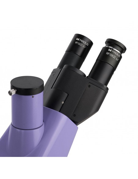
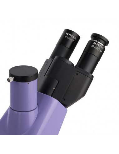
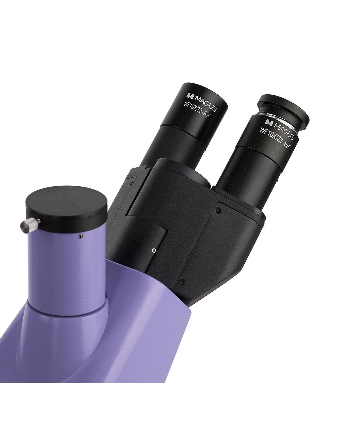
This microscope is equipped with a classic trinocular head, whose vertical tube is used to house a digital camera. The camera is not included in the package. The beam splitter along the optical path has a 50/50 ratio.
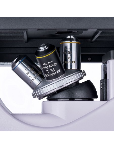
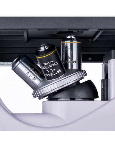
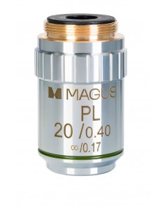
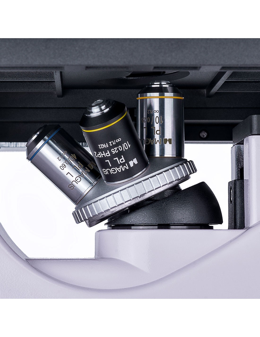
Four included objectives or others purchased separately can be housed on the nosepiece. All included objectives are designed for use with laboratory glassware and have a long working distance. Specifically, 3 planachromatic objectives for bright field observations and 1 planachromatic objective for phase contrast microscopy are included.
Illumination
The light source for transmitted light observations it is a 9 W LED. The high intensity of the LED ensures that the illumination of the field of view is sufficient to work with all objectives and with each of the expected microscopy techniques. Adjusting the brightness does not cause any change in color temperature. The life cycle of the LEDs is approximately 50,000 hours without any bulb replacement.
The capacitor has an available slot for a phase contrast slider (included). You can center the phase contrast rings. The inclusion of the slider allows you to save time when you want to switch from one microscopy technique to another.
Stage and focusing mechanism
Fixed translating table. It is equipped with plate holders (three slots for different sized glassware are included). The plates can be moved along the X-axis and Y-axis using a translator insert. The focusing mechanism with coaxial macro and micrometric screws allows you to focus the image quickly and accurately. The coarse focus (coarse screw) is equipped with a fixing mechanism and the tension of the coarse screw can be adjusted.
Accessories
The line of optional accessories includes lenses, eyepieces, digital cameras and calibration slides.
Features microscope key:
- Bright field and phase contrast microscopy techniques in transmitted light; study of samples in laboratory glassware
- Digital camera installed in the trinocular tube
- 9 W LED illuminator for transmitted light observations with adjustable brightness and long life cycle
- Fixed stage with movement along the two axes, 3 plate holders of different sizes included
- Optional accessories to improve the performance of the microscope
Key features of the camera:
- Back-illuminated color SONY Exmor CMOS sensor, highly sensitive to light
- Dark and bright field operation via 4x, 10x and 20x objectives
- Connection to a computer via a USB 3.0 interface with a data transfer rate of 5 Gbps
- Image reproduced on a monitor at 4K Ultra High resolution 3840x2160 px (45 fps) or Full HD 1920x1080 px (70 fps)
- Driver and image processing software are included
- Installation on the trinocular tube or optical tube of a microscope
The package contains:
- MAGUS CDF30 digital camera (digital camera, USB cable, installation CD with drivers and software, user manual and warranty card)
- Support with integrated power supply, transmitted light source, focusing mechanism, stage, condenser with housing for phase contrast slide and rotating objective holder
- Trinocular head
- Infinity plan achromatic objective: PL 10x/0.25 WD 4.27 mm
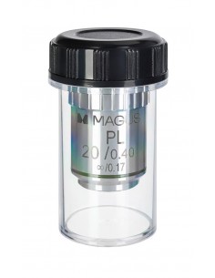
- Infinity plan achromatic objective: PL L20х/0.40 WD 8.0 mm
- Infinity plan achromatic objective: PL L40х/0.60 WD: 3.5 mm
- Infinity plan achromatic objective: PHP2 10x/0.25 phase contrast WD 4.27 mm
- 10x/22 mm eyepiece with long eye relief (2 pcs.)
- Eyecup (2 pcs.)
- Centering telescope
- Center slider phase contrast with centering phase contrast rings
- Mechanical translator for moving samples
- Plate support (3 pcs.)
- C-pitch adapter
- Color filter
- AC power cord
- Dust cover
- User manual and warranty card ul>
Available on request:
- 10x/22 mm eyepiece with graduated scale
- 12.5x/14 mm eyepiece (2 pcs.)
- Eyepiece 15x/15 mm (2 pcs.)
- Eyepiece 20x/12 mm (2 pcs.)
- Eyepiece 25x/9 mm ( 2 pcs.)
- Infinity plan achromatic objective: PL 4x/0.10 WD 21 mm
- Infinity plan achromatic objective: PHP2 20x/0.40 phase contrast WD 8 .0 mm
- Infinity plan achromatic objective: PHP2 40x/0.60 phase contrast WD 3.5 mm
- Calibration slide
- LCD monitor
| Brand | MAGUS |
| Warranty | 5 лет |
| EAN | 5905555018096 |
| Package size (WxDxH) | 64x42x67 cm |
| Shipping weight | 18.6 kg |
| Delimiter | MAGUS Bio V300 Inverted Biological Microscope |
| === Delimiter === | Microscope Specifications |
| Type | biological, light/optical, digital |
| Tested | trinocular |
| Vertical tube | Siedentopf |
| Head inclination angle | 45° |
| Magnification, x | 100–400 in basic configuration (*optional: 40–500/600/800/1000) |
| Range magnification | from 200x to 800x |
| Eyepiece tube diameter, mm | 30 |
| Eyepieces | 10х/22 mm, eye relief: 10 mm (*optional: 10x/22 mm with graduated scale, 12.5x/14; 15x/15; 20x/12; 25x/9) |
| Infinity planachromatic | objectives: PL 10x/0.25; PL 20x/0.40; PL 40x/0.60; PHP2 10x/0.25 phase; parfocal distance: 45 mm (*optional: PL 4x/0.10; PHP2 20x/0.40 phase; PHP2 40x/0.60 phase) |
| Nosepiece nosepiece |
| Nosepiece | for 4 objectives |
| Working distance, mm | 4.27 (10x); 8.0 (20х); 3.5 (40x) |
| Interpupillary distance, mm | 48 — 75 |
| Range of stage movement, mm | 77/112 |
| Stage features | fixed, with mechanical translator for moving the sample; plate holder: 29x77 mm, Ø90 mm; 34x77.5 mm, Ø68.5 mm; 57x82 mm, Ø60 mm |
| Condenser | NA0.6, working distance: 70 mm; with adjustable aperture diaphragm and a housing for phase contrast slider |
| Diaphragm | adjustable aperture |
| coaxial, coarse focus (with micrometer screw tension adjustment knob and fixing knob) and fine focus (0.002 mm micrometer screw) |
| Lighting | LED |
| Brightness adjustment | yes |
| Power supply | 220±22 V, 50 Hz, AC mains |
| Type of light source | 9 W |
| Optical filters | yes |
| Operating temperature range, °C |
| Special Features | phase contrast slider for 10x objectives with a centrable phase contrast ring |
| User level | expert users, professionals |
| Assembly and installation level of difficulty | complicated< /td> |
| Application | laboratory/medical |
| Lighting position | lower |
| td> |
| Research method | bright field, phase contrast microscope |
| Set with case/case/ bag | dust cover |
| === Delimiter === | Digital camera |
| Sensor | SONY Exmor CMOS |
| Color/monochrome | color |
| Megapixel | 8.3 |
| Maximum resolution, px | 3840x2160 |
| Sensor size | 1/1.2'' (11.14x6.26 mm) |
| Pixel size, µm | 2.9x2.9 |
| Light sensitivity | 5970 mV/lux/s |
| Signal-to-noise ratio | 0.13 mV with 1/30 s |
| Exposure time | 0.02 ms – 15 s |
| Video recording | yes |
| Video recording | yes |
| Frame rate, fps@resolution | 45@3840x2160, 70@1920x1080 |
| Installation location | trinocular tube, optical tube instead of eyepiece |
| Image format | *.jpg, *.bmp, * .png, *.tif |
| Video format | recording: *.wmv, *.avi, *.h264 (Windows 8 and later), * h265 (Windows 10 and later) |
| Spectral range, nm | 380-650 (integrated IR filter) |
| Shutter type | ERS (electronic curtain shutter) |
| White balance | manual, automatic |
| Exposure control | manual, automatic |
| Software features | image size, brightness, exposure control |
| Software | MAGUS View |
| Output |
| System requirements | Windows 8/10/11 (32 bit and 64 bit), Mac OS , Linux, up to 2.8 GHz Intel Core 2 or later, minimum 2 GB RAM, USB 3.0 port, CD-ROM, 17" or larger |
| Type of mount | C-mount |
| Body | robust aluminum |
| Camera power supply | DC, 5 V, from computer USB port; 12 V, 3 A adapter for the Peltier cell |
| Camera operating temperature range, °C | -10 — 50 |
| Humidity operating range, % | 30 — 80 |