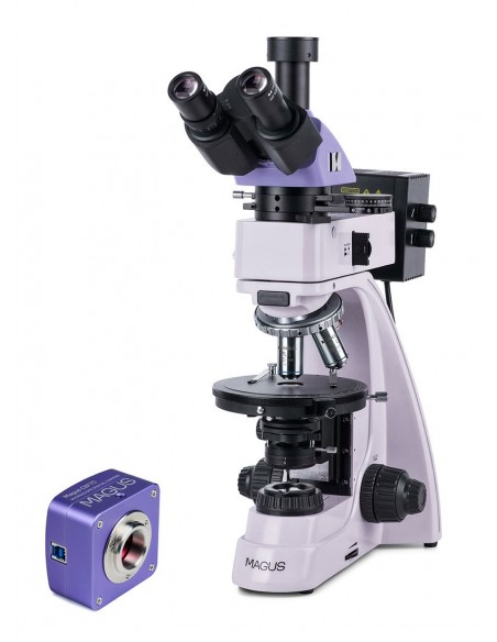
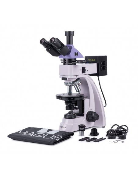
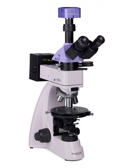
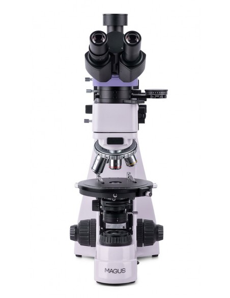
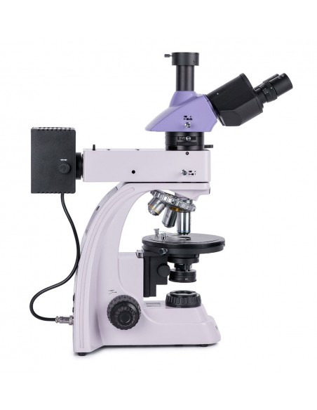
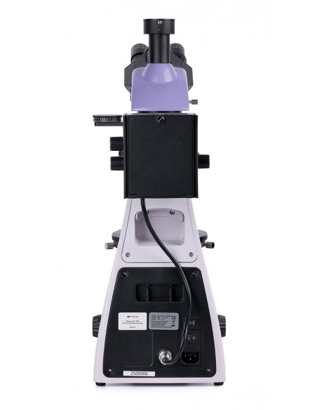
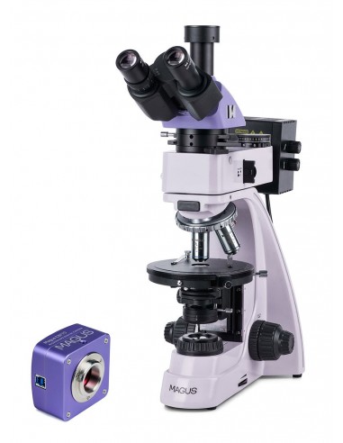
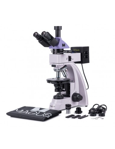
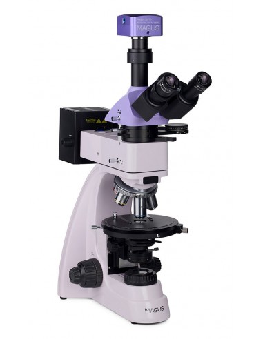
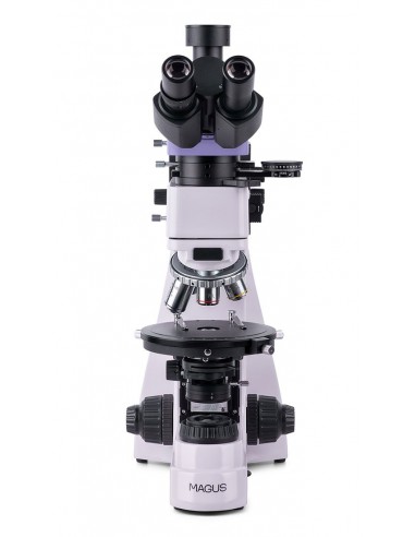
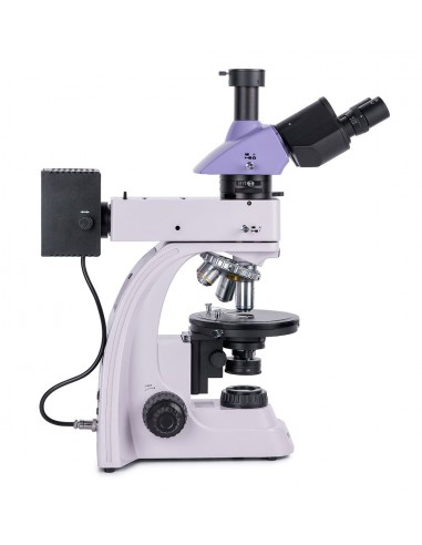
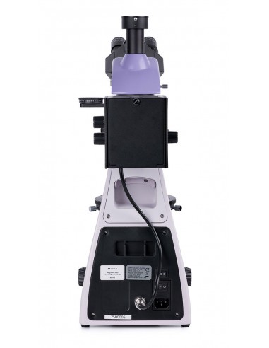
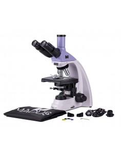
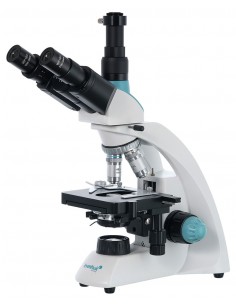
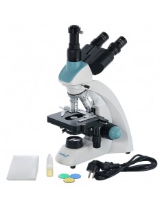
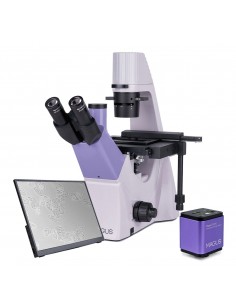
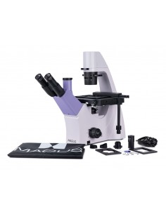
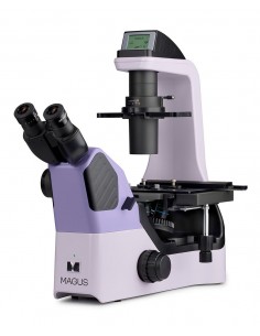
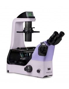
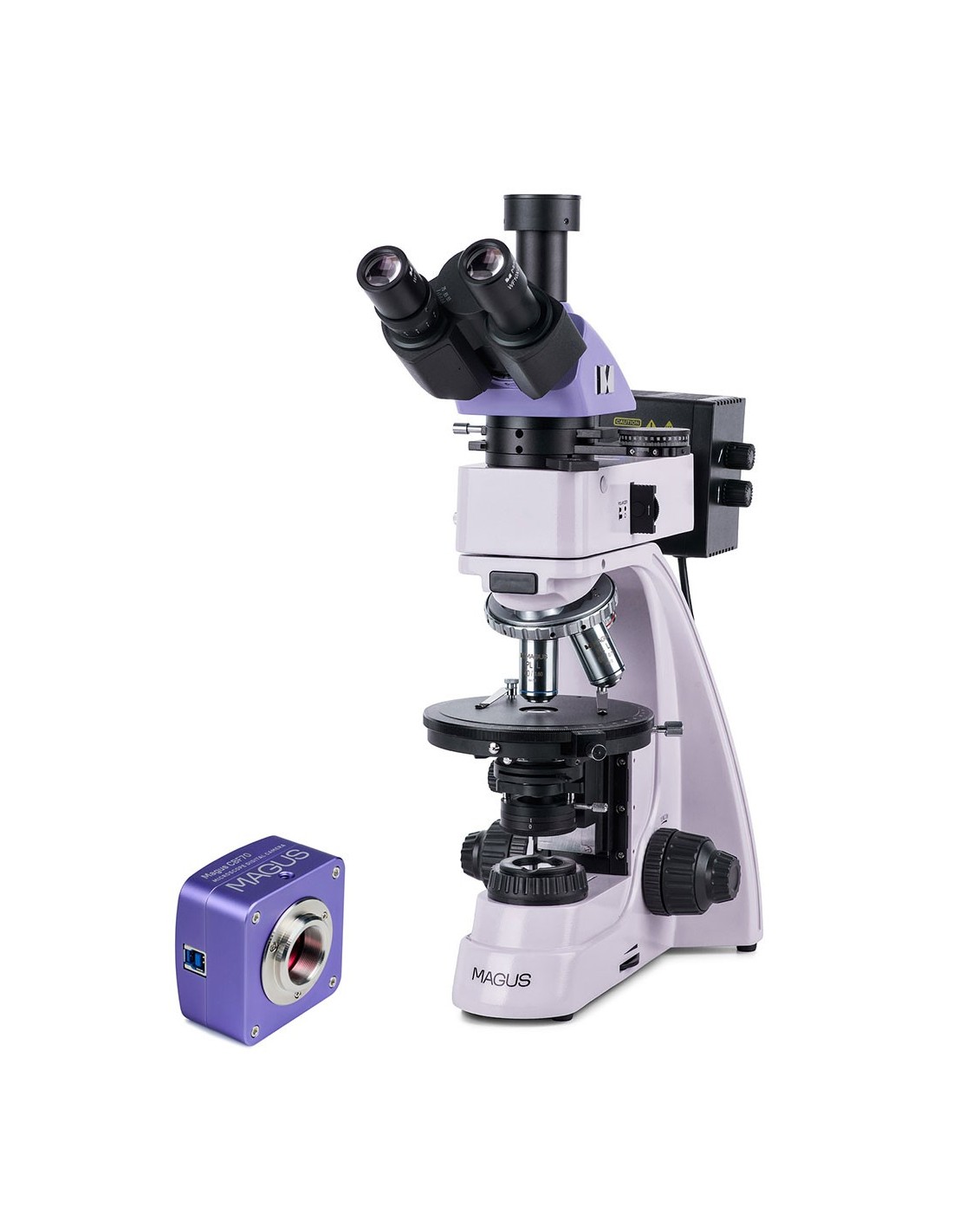
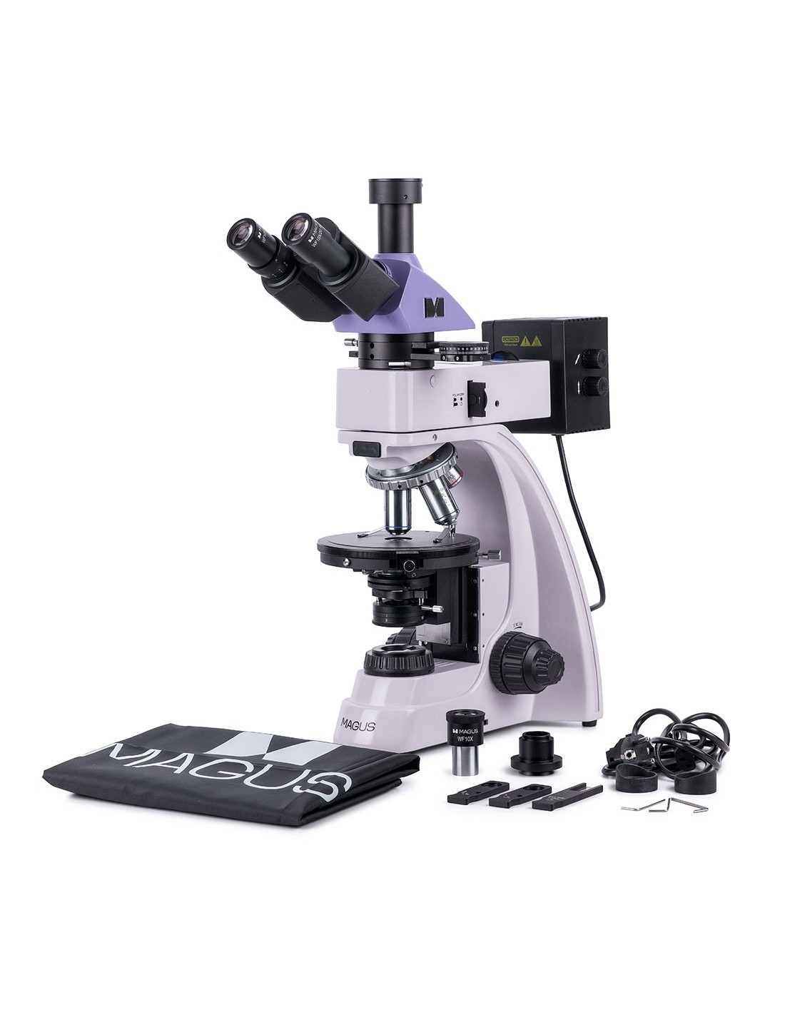
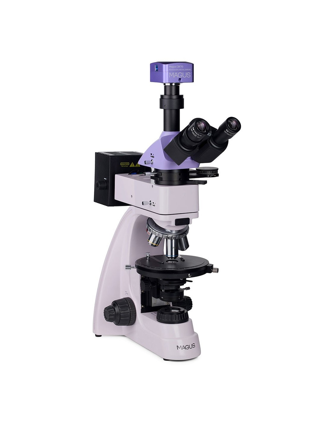
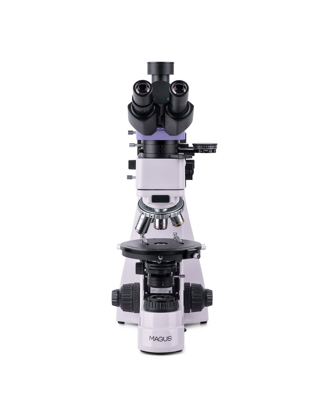
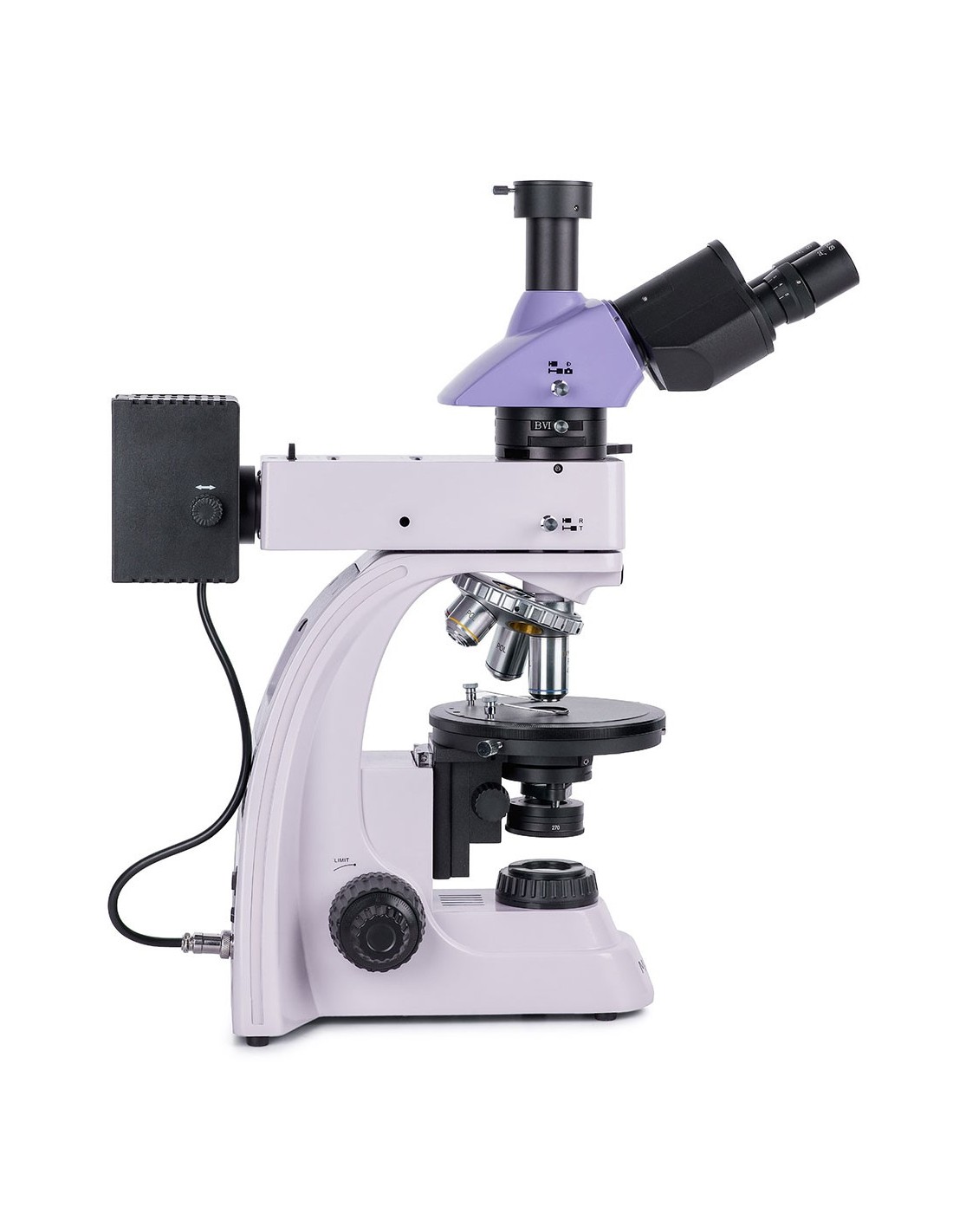
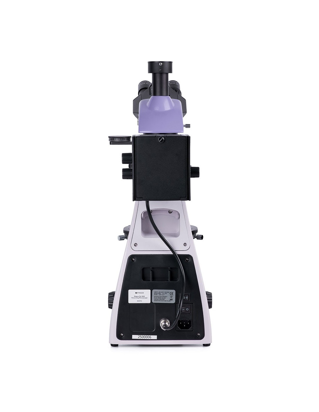
The MAGUS Pol 850 polarizing microscope is designed to operate in transmitted and reflected light. Available research methods: polarization and bright field. The microscope is used to study anisotropic anibiological, geological and polymeric objects, as well as opaque polished sections up to 15 mm thick with a polished side. The microscope is perfect for a wide range of tasks of professionals in the fields of medicine, forensics, mineralogy, crystallography or petrography.
Digital camera
The MAGUS CBF70 professional scientific camera is designed for use with a microscope as well as 4x, 10x, 20x and 40x objectives. The device allows you to take photographs, shoot videos, organize demonstrations, display images on an external screen and perform angular and linear measurements of study objects (for this function to be performed correctly, the software must be calibrated). The camera is recommended for working with the bright field method.
The MAGUS CBF70 camera has five image resolution and frame rate modes, so among them you can easily find the one that best suits your needs. your needs: 5280x3954 px (17 fps), 3952x3952 px (17 fps), 2640x1976 px (56 fps), 1760x1316 px (67 fps) and 584x438 px (192 fps).
The high resolution modes are ideal for photography, allowing you to create clear, vibrant and highly detailed images. Modes with increased frame rate will help when recording smooth and informative video, without jumps and delays, recording the smallest movements of the object or features of fast processes observed.
To obtain the best image quality without distortions , it is recommended to use an adapter with 1x magnification.
The camera design uses a 4/3” color back-illuminated SONY Exmor CMOS sensor. It makes the image bright even in a low-light environment. The larger physical size of the sensor helps the colors displayed appear deeper and the image appear clearer.
Optics
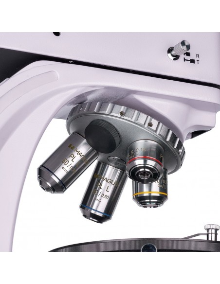
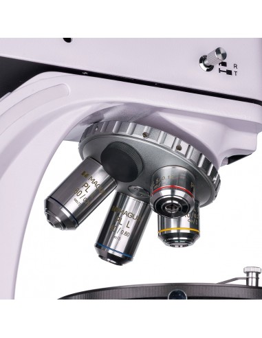
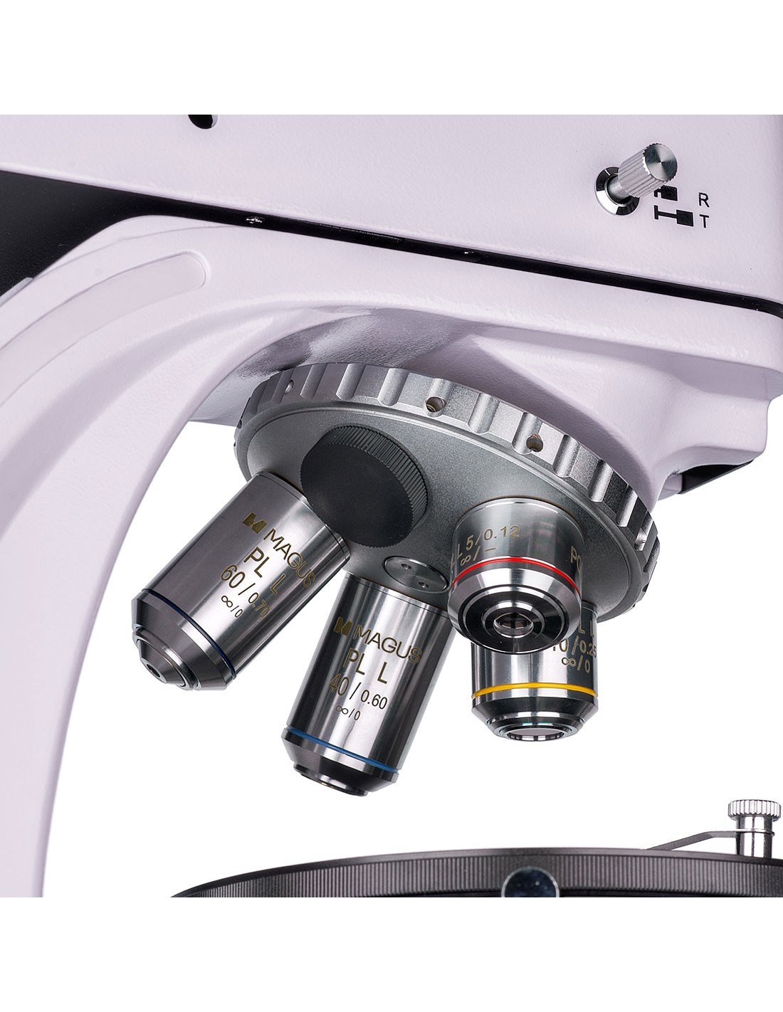
The microscope's optics are corrected to infinity and are free from internal tensions. The image formed by the lens is authentic, glare-free, high-contrast and detailed. The lens is installed in a nosepiece with grooves centered on the optical axis. There are five grooves, with four lenses included in the kit; An additional objective can be installed to achieve further magnification within the magnification range of the microscope. The objectives are designed to work with specimens without coverslips.
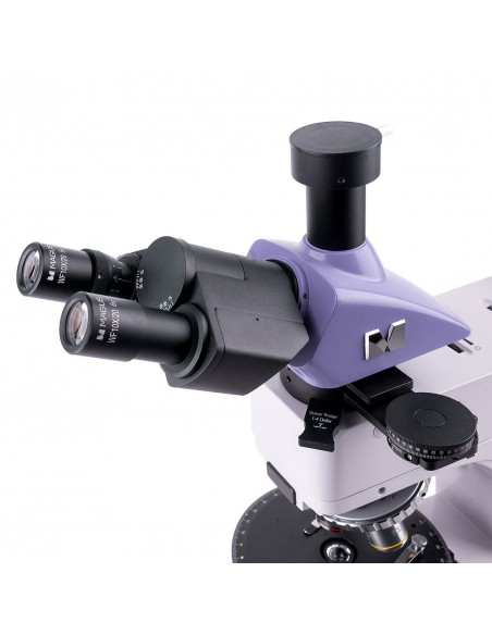
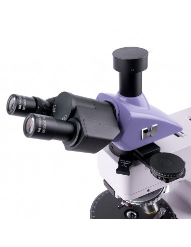
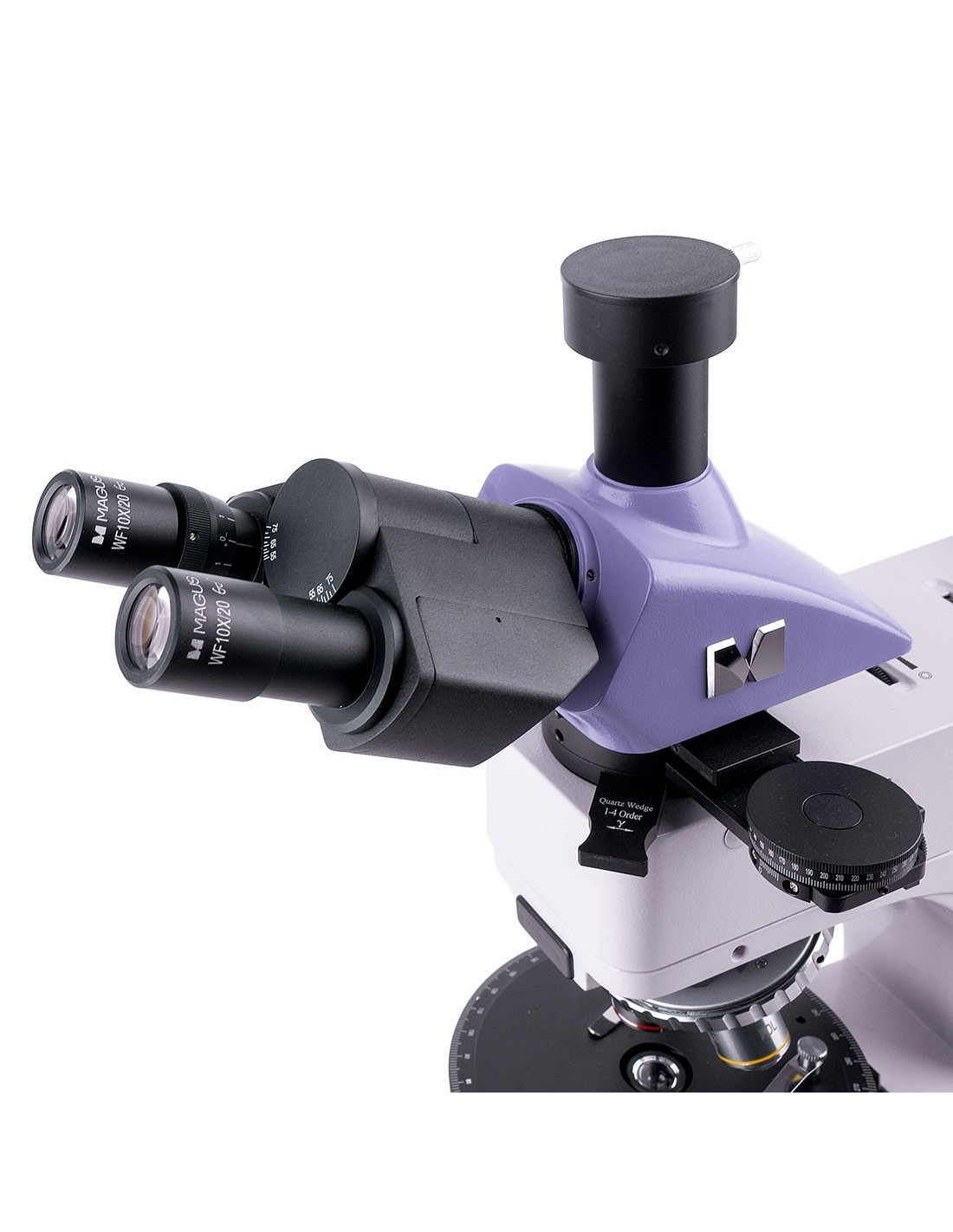
The trinocular head allows you to install a digital camera in the trinocular tube. The beam split is 100:0 or 0:100, meaning all the light goes to the camera or eyepiece tubes. The left eyepiece tube is equipped with a diopter adjustment ring: it is rotated to compensate for the difference in vision between the eyes and to obtain the clearest possible image of the specimen.
Illumination
strong>
30W halogen lamps are used for transmitted and reflected light. Their brightness is sufficient to obtain a clearly readable image on each lens, and the “warm” emission spectrum is comfortable during long-term use. The Köhler illumination setting will provide greater resolution and remove artifacts in the field of view and darkening at the edges. Light filters are used to fine-tune color reproduction when working in reflected light.
Polarization is available for both transmitted and reflected light. To begin observations in polarized light, an analyzer is introduced into the optical path. Next, the relative position of the analyzer and polarizer is changed by rotating them, which also changes the polarization angle. The contrast of objects is increased with the help of compensators installed in a special housing in the intermediate tube. The same attachment also contains a Bertrand lens which is used for Conopic examinations.
Stage and focusing mechanism
The microscope has a rotatable stage: rotating it changes how the sample refracts light in the presence of polarized light. The stage is equipped with a scale on which the angle of rotation can be determined with an accuracy of up to 0.1°. To get the brightest and clearest image, the stage can be centered on the optical axis of the microscope.
The coarse and fine focus knobs are located at the bottom of the stand, so the user can assume a relaxed pose with hands on the table. The coarse tension can be adjusted and there is also a coarse focus lock. The fine focus ensures smooth movement of the sample.
Accessories
To make the most of the microscope, use accessories from the MAGUS line: objective eyepieces, calibration slides, digital cameras, etc.
Key features of the microscope:
- For work in transmitted, reflected polarized and polarized light conditions natural
- Trinocular head with digital camera tube; Beam split of 100:0 or 0:100
- Voltage-free planachromatic objectives for sharp images free from artifacts and false internal reflections
- Halogen illumination of the working area, a spectrum of colors comfortable for the eyes; lamp power 30 W
- Rotating stage, polarizer and analyzer with rotation angle markings, Bertrand lens and compensators
- The grooves of the revolver and the stage are centered on the axis microscope optical
- Compatible with additional accessories
Key camera features:
- Five graphics settings and fps: between them you can easily find the most suitable mode
- Fast data transfer thanks to the USB 3.0 interface
- Large SONY Exmor color sensor for bright and clear images even in low light conditions
- “Quick start”: install the included software and then the camera is ready for use
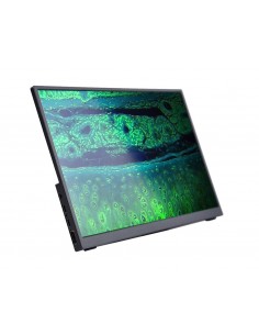
- Easy observations or demonstrate to an audience by displaying the camera image on an external screen
The package contains:
- MAGUS CBF70 digital camera (digital camera, USB cable , installation CD with drivers and software, user manual and warranty card)
- Base with power input, transmitted light light source and condenser, focusing mechanism, stage and nosepiece
- Illuminator for reflected light observations with bulb housing
- Trinocular head
- Intermediate tube with Bertrand lenses, analyzer and compensator housing
- Compensators: λ compensator, λ/4 compensator, quartz wedge
- Infinity plan achromatic objective: PL 5x/0.12 WD 26.1 mm
- Infinity plan achromatic objective: PL 10x /0.25 WD 5.0 mm
- Infinity plan achromatic objective: PL 40x/0.6 (with spring mechanism) WD 3.98 mm
- Infinity plan achromatic objective infinity: PL 60x/0.7 (with spring mechanism) WD 2.03 mm
- Eyepiece 10x/20 mm (2 pcs.)
- Eyepiece 10x/20 mm with scale graduated. Scale value: 0.1 mm
- 1x C-mount adapter
- Allen key
- Power cable
- Dust cover
- Dust cover li>
- User manual and warranty certificate
Available on request:
- 16x/11 mm eyepiece (2 pcs.)
- Eyepiece 20x/11 mm (2 pcs.)
- Infinity planachromatic objective: PL 2.5x/0.07 WD 11 mm
- Infinity plan achromatic objective: PL 50x/0.7 (with spring mechanism) WD 3.67 mm
- Infinity plan achromatic objective: PL 80x/0.8 (with spring mechanism) WD 1.25 mm
- Planachromatic infinity objective: PL 100x/0.85 (with spring mechanism) WD 0.4 mm
- Mechanical stage
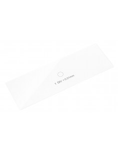
- Calibration glass
| Brand | MAGUS |
| Warranty | 5 лет |
| EAN | 5905555018546 |
| Package size (LxWxH) |
| td> | 44x42x67 cm |
| Shipping weight | 12.1 kg |
| Delimiter | MAGUS Pol 850 polarizing microscope |
| === Delimiter === | Microscope specifications |
| Type | biological, light/optical, digital |
| Tested | trinocular |
| Vertical tube | Siedentopf |
| Head inclination angle | 30° |
| Magnification, x | 50–600 in basic configuration (*optional: 25–1000/1600/2000) |
| Eyepiece tube diameter, mm | 23.2 |
| Eyepieces | 10х/20; 10x/20 with graduated scale (*optional: 16x/11; 20х/11) |
| Infinity planachromatic objectives, non-deforming: PL 5x /0.12; PL 10x/0.25; PL 40x/0.6; PL 60x/0.7 (*optional: PL 2.5x/0.07; PL 50x/0.7; PL 80x/0.8; PL 100x/0.85); parfocal height of 45 mm, designed for the study of samples without coverslips |
| Nosepiece nosepiece | for 5 objectives, centering |
| Working distance, mm | 26.1 (5x); 5.0 (10x); 3.98 (40x); 2.03 (60x); 11 (2.5x); 3.67 (50x); 1.25 (80x); 0.4 (100x) |
| Stage, mm | Ø150 |
| Stage features | 360° rotatable, centering, gradation equal to 1° of the rotation angle; a vernier scale for measuring angles with an accuracy of 0.1° |
| Diopter adjustment of the eyepiece, diopters | ±5 (on the barrel left) |
| Condenser | Abbe NA 1.25 condenser with an adjustable aperture diaphragm and rotatable lenses |
| Diaphragm | adjustable aperture diaphragm, field diaphragm with adjustable iris |
| Focus | coaxial, coarse focus (21 mm, 39.8 mm/turn, with fixing knob and micrometer screw tension adjustment knob) and fine focus (0.002 mm micrometer screw) |
| Lighting | halogen |
| Brightness adjustment | yes |
| Power | 220±22 V, 50 Hz, AC mains |
| Type of light source | reflected and transmitted light: 12 V/ 30 W |
| Optical filters | reflected light: opaque, yellow, green, blue |
| Operating temperature range, °C | 5 — 35 |
| User level | expert users, professionals |
| Assembly and installation level of difficulty | complicated |
| Polarizer | transmitted light: with 0 °, 90°, 180°, 270° marked on the scale; 360° rotatable; with reflected light: removable |
| Intermediate tube | integrated analyzer rotatable from 0–360°; a vernier scale for measuring angles with an accuracy of 0.1°; Bertrand lenses; housings for installing compensators |
| Compensator | compensator λ; λ/4 compensator; quartz wedge |
| Application | laboratory/medical |
| Lighting position | double |
| Research method | bright field, polarized light |
| Set with case/case /bag | dust cover |
| === Boundary === | Digital camera |
| Sensor | SONY Exmor CMOS |
| Color/monochrome | color |
| Maximum resolution, px | 5280x3954 |
| Sensor size | 4/3" (17, 4x13.0 mm) |
| Pixel size, µm | 3.3x3.3 |
| Sensitivity to light | 399 mV at 1/30 s |
| Signal to noise ratio | 0.1 mV at 1/30 s< /td> |
| Exposure time | 0.1 ms – 15 s |
| Video recording | yes |
| Video recording | yes |
| Frame rate, fps@resolution< /td> | 17@5280x3954, 192@584x438, 67@1760x1316, 56@2640x1976, 17@3952x3952 |
| Installation location | tube trinocular, optical tube instead of eyepiece |
| Image format | *.jpg, *.bmp, *.png, *.tif |
| Video format | recording: *.wmv, *.avi, *.h264 (Windows 8 and later), *h265 (Windows 10 and later) |
| Spectral range, nm | 380-650 (integrated IR filter) |
| Shutter type |
| White balance | manual, automatic |
| Exposure control | manual, automatic |
| Software features | image size, brightness, exposure control |
| Software | MAGUS View |
| Output | USB 3.0, 5 Gb/s |
| System requirements | Windows 8/10/11 (32 bit and 64 bit), Mac OS or later, minimum 2 GB RAM, USB 3.0 port, CD-ROM, 17" or larger |
| Mount type | C-mount |
| td> |
| Body | robust aluminum |
| Camera power supply | DC, 5 V, from the computer's USB port; 12 V, 3 A adapter for the Peltier cell |
| Camera operating temperature range, °C | -10 — 50 |
| Humidity operating range, % | 30 — 80 |